
Shoulder Impingement Syndrome is one of the most common causes of shoulder pain and restricted movement, and can progress to a partial or complete rotator cuff tear if left untreated.
Treatment Goal
The goal of treatment is to reduce pain and restore normal shoulder function. A shoulder tendon specialist designs the treatment plan based on the patient’s age, activity level, and general health.
Causes of Subacromial Impingement
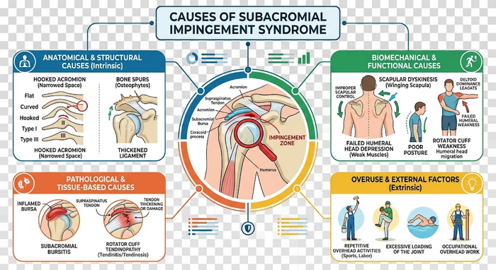
Subacromial impingement syndrome results from compression and inflammation of the rotator cuff tendons and subacromial bursa within the subacromial space. Causes are divided into intrinsic and extrinsic:
Intrinsic Causes
- Rotator cuff muscle weakness, causing the humeral head to migrate superiorly.
- Repetitive overuse causing tendinitis and subacromial bursitis.
- Degenerative changes in the rotator cuff tendons, reducing available space.
Extrinsic Causes
- Acromion shape variants — a hooked (Type III) acromion directly compresses the tendon during arm elevation.
- Shoulder muscle weakness or dyskinesia affecting humeral head stability.
- Glenohumeral instability causing dynamic superior migration of the humeral head.
- Acromioclavicular joint osteophytes that narrow the subacromial corridor.
Overhead athletes (swimmers, handball, volleyball players) and workers who repeatedly lift their arms above shoulder height are at highest risk.
Causes Classification — Internal vs. External
| Cause | Type | Mechanism | Most Affected Group | Modifiable? |
|---|---|---|---|---|
| Rotator cuff muscle weakness | Intrinsic | Superior humeral migration → tendon compression | Athletes, desk workers | Yes — strengthening programme |
| Repetitive overuse | Intrinsic | Micro-tears → chronic tendon inflammation | Overhead athletes, swimmers | Yes — training modification |
| Rotator cuff tendon degeneration | Intrinsic | Progressive tendon weakening → narrowed space + tear risk | Over 40 | Partial — exercise and injections |
| Type III (hooked) acromion | Extrinsic | Hook directly compresses tendon on arm elevation | Hereditary — any age | No — surgery if symptomatic |
| AC joint osteophytes | Extrinsic | Bony spurs narrow subacromial corridor | Over 50 | No — surgical removal required |
| Glenohumeral instability | Extrinsic | Dynamic superior humeral migration | Young throwing athletes | Yes — stabilisation and physio |
| * Based on AAOS, Mayo Clinic, and ASES guidelines. Most cases involve more than one cause. Identifying the primary driver is essential for optimal treatment selection. | ||||
Diagnostic Tools Comparison
| Tool | What It Shows | What It Misses | Rotator Cuff Accuracy | Relative Cost | First-Line Use |
|---|---|---|---|---|---|
| X-ray | Osteophytes, calcifications, subacromial space, OA | Tendons and soft tissue | Does not image tendons | Low | Always — first step |
| Ultrasound | Bursal fluid, bursa wall thickening, partial surface tear | Deep tears — does not assess bone | Good for partial surface tears | Low–moderate | Before MRI; also guides injections |
| MRI | Full / partial tears, bursitis, degeneration, fluid accumulation | Dynamic tendon assessment | Very high — gold standard | High | Suspected tear; before surgery |
| Hawkins-Kennedy test (clinical) | Active subacromial impingement | Does not localise or grade injury | Sensitivity 79%, Specificity 59% | No cost | Routine clinical examination |
| Neer test (clinical) | Impingement on arm elevation | Does not differentiate impingement from tear | Sensitivity 72%, Specificity 60% | No cost | Routine clinical examination |
| * Clinical test sensitivity from Hegedus et al. BJSM 2008 systematic review. MRI is the gold standard for rotator cuff tendon assessment before surgical decision-making. | |||||
Does Every Patient Need Surgery?
No. Most cases begin with non-surgical treatment — rest, medication, and physiotherapy — with most patients experiencing progressive improvement over weeks to months.
Non-Surgical Treatment
- Rest and activity modification: Avoid overhead movements and contact sports involving the shoulder.
- NSAIDs: Ibuprofen, naproxen or aspirin to reduce pain and swelling.
- Physiotherapy: Range-of-motion exercises, rotator cuff strengthening, and posture correction.
- Corticosteroid injection: If symptoms persist, a subacromial cortisone injection rapidly reduces inflammation.

Conservative vs. Surgical Treatment — Comparison
| Criterion | Conservative Treatment | Arthroscopic Surgery (Subacromial Decompression) |
|---|---|---|
| Ideal candidate | Impingement < 6 months — no complete tear | Failed conservative > 3–6 months — impingement with tear or osteophytes |
| Pain relief success rate | 60–80% at 3–6 months | 80–90% |
| Return to desk work | Weeks (with activity modification) | 2–4 weeks |
| Return to overhead sport | 3–6 months | 4–6 months |
| Risk of progression to complete tear if neglected | High with neglect | Removes the structural cause of tearing |
| Full rehabilitation period | 6–12 weeks | 3–6 months for full recovery |
| * Rates from AAOS, Mayo Clinic, and AJSM. Surgical decision is made by the treating surgeon after examination and imaging. | ||
Shoulder Arthroscopy Cost Calculator
Estimated surgical expenditure in Egypt (Private & Academic Centers)
Post-Surgical Rehabilitation Protocol
| Phase | Weeks | Goals | Recommended Exercises | Permitted Activities | Restricted Activities |
|---|---|---|---|---|---|
| Acute | 1–2 | Pain and swelling control — protect repair | Pendulum exercises, limited passive ROM | Simple ADLs with sling | Overhead reaching — any lifting |
| Range-of-Motion Restoration | 3–6 | Restore full passive and active ROM | Active stretching — progressive shoulder arc | Desk work — driving (with clearance) | Weight bearing — contact sports |
| Strengthening | 7–12 | Rotator cuff and shoulder stabiliser strength | Resistance band — deltoid and rotator cuff exercises | Light swimming — non-shoulder sports | Throwing — overhead sports |
| Return to Activity | 13–24+ | Full sport and occupational return | Functional training — full shoulder mechanics | All activities progressively | Competitive sport without physician clearance |
| * Protocol applies to isolated subacromial decompression. If a concurrent rotator cuff repair was performed, the protection phase extends to 6 weeks. All rehabilitation must be supervised by a qualified physiotherapist under the treating surgeon’s guidance. | |||||
Frequently Asked Questions About Shoulder Impingement Syndrome
Evidence-based insights into causes, diagnostic maneuvers, non-surgical therapies, and arthroscopic management.
What is Shoulder Impingement Syndrome (Subacromial Impingement)?
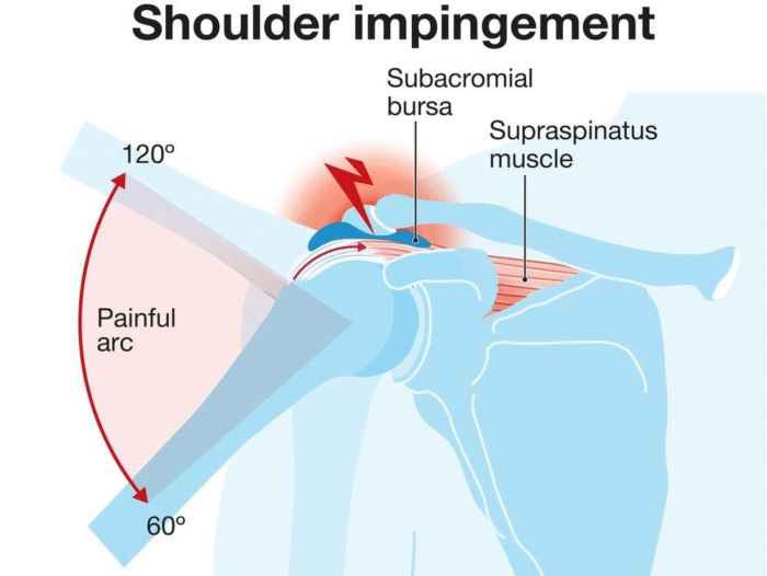
Shoulder Impingement Syndrome (SIS), clinically referred to as Subacromial Impingement Syndrome (SAIS) or Subacromial Pain Syndrome (SAPS), is a musculoskeletal condition characterized by mechanical irritation and compression of the rotator cuff tendons (primarily the supraspinatus) and the subacromial bursa as they pass through the narrow subacromial space beneath the coracoacromial arch during arm elevation.
What are the hallmark clinical symptoms of shoulder impingement?
Patients classically report an anterior-lateral shoulder ache radiating to the lateral deltoid insertion. Key indicators include:
• A “painful arc” between 60° and 120° of active arm elevation.
• Nocturnal pain exacerbated by lying on the affected side.
• Discomfort when reaching overhead, behind the back (e.g., reaching for a back pocket), or during sports involving throwing and swimming.
What is the difference between primary and secondary impingement?
Primary impingement is an anatomical or structural outlet narrowing caused by a hooked acromion (Bigliani Type III), subacromial bone spurs, or hypertrophic degenerative changes of the acromioclavicular (AC) joint.
Secondary impingement is dynamic or functional; the subacromial space narrows due to glenohumeral capsular laxity, scapular dyskinesis, or rotator cuff muscle fatigue, allowing the humeral head to migrate superiorly during active elevation.
How is shoulder impingement diagnosed by an orthopedic surgeon?
Diagnosis begins with specialized clinical physical examination tests (Neer sign, Hawkins-Kennedy test, Jobe’s empty can test). Imaging includes standard radiographs (True AP, Axillary, and Scapular Y / Outlet views) to assess acromial morphology and calcific deposits, while high-resolution MRI or musculoskeletal ultrasound is utilized to assess tendon integrity, bursal thickening, and rule out full-thickness rotator cuff tears.
What is the Neer test and the Neer diagnostic injection test?
The Neer test is a physical maneuver where the examiner prevents scapular rotation while passively flexing the patient’s internally rotated arm forward, reproducing impingement pain under the anterior acromion.
The Neer injection test involves injecting 5 to 10 mL of 1% lidocaine into the subacromial space. If the patient experiences immediate pain relief upon repeating provocative maneuvers, the diagnosis of subacromial pathology is confirmed.
What is the first-line non-surgical treatment protocol?
Conservative management is successful in up to 70–80% of patients and is the gold standard for initial care:
• Activity Modification: Avoiding exacerbating overhead motions.
• Medication: Short course of oral non-steroidal anti-inflammatory drugs (NSAIDs) for pain control.
• Targeted Physical Therapy: Eccentric strengthening of the rotator cuff (infraspinatus, teres minor, subscapularis) to depress the humeral head, alongside periscapular stabilization (serratus anterior and lower trapezius) to correct scapular rhythm.
Are subacromial cortisone injections safe and effective?
Subacromial corticosteroid injections provide powerful short-term relief from acute subacromial bursitis and can facilitate participation in physical rehabilitation. However, repeated injections are not advised due to potential adverse effects on tendon histology, including impaired tenocyte collagen synthesis and increased risk of tendon rupture.
Can untreated shoulder impingement lead to a rotator cuff tear?
Yes. Charles Neer described three progressive stages of impingement syndrome:
• Stage I: Reversible edema and hemorrhage (usually in young active individuals < 25 years).
• Stage II: Fibrosis and tendinitis of the rotator cuff tendon (typically aged 25–40).
• Stage III: Formation of bone spurs and partial or complete tears of the rotator cuff and biceps tendon.
When is surgery recommended for shoulder impingement?
Surgery is considered when a compliant patient experiences persistent or worsening symptoms after 3 to 6 months of supervised non-operative therapy, when mechanical blockades (large inferior acromial spurs) impede motion, or when full-thickness rotator cuff tears develop requiring concomitant repair.
What surgical procedure is performed, and how does it work?
The standard surgical procedure is Arthroscopic Subacromial Decompression (ASD) with acromioplasty. Through 2 to 3 keyhole incisions (approx. 5 mm each), the surgeon utilizes an arthroscope and motorized burr to:
1. Remove the inflamed, thickened subacromial bursa (subacromial bursectomy).
2. Release the coracoacromial ligament.
3. Shave and flatten the protruding anterior and inferior border of the acromion to create a smooth, expanded corridor for tendon excursion.
References
- AAOS OrthoInfo. Rotator Cuff Tears. 2023. Available at: orthoinfo.aaos.org
- AAOS OrthoInfo. Shoulder Impingement/Rotator Cuff Tendinitis. 2023. Available at: orthoinfo.aaos.org
- Mayo Clinic Staff. Rotator cuff injury – Symptoms and causes. 2023. Available at: mayoclinic.org
- Cleveland Clinic. Shoulder Impingement Syndrome. 2023. Available at: my.clevelandclinic.org
- Hospital for Special Surgery (HSS). Shoulder Impingement and Rotator Cuff Disease. 2023. Available at: hss.edu
- Hegedus EJ, et al. Physical examination tests of the shoulder: a systematic review with meta-analysis. British Journal of Sports Medicine. 2008;42(2):80–92. DOI: 10.1136/bjsm.2007.038406
- Papadonikolakis A, et al. Published evidence for shoulder surgery: a systematic review. Journal of Bone and Joint Surgery. 2011;93(22):577–585. Available at: ncbi.nlm.nih.gov
- Medscape. Shoulder Impingement Syndrome Treatment & Management. 2024. Available at: emedicine.medscape.com