
Do you experience sharp, nagging pain at the back of your heel — especially when you first step out of bed in the morning or after exercise? This pain may be more than temporary strain. It is often a sign of Achilles tendonitis, one of the most common musculoskeletal injuries affecting athletes and non-athletes alike.
Ignoring this pain can lead to worsening damage and potentially serious complications, including complete tendon rupture that may require surgical repair.
In this comprehensive guide, we cover everything you need to know about the Achilles tendon — from anatomy and causes of inflammation to the best available treatments for Achilles tendonitis.
What Is the Achilles Tendon?
The Achilles tendon is not just any tendon — it is the strongest and largest tendon in the human body. It is a powerful fibrous connective tissue that functions like a thick cord, connecting the posterior calf muscles (the gastrocnemius and soleus) to the heel bone (calcaneus). This connection gives you the ability to stand on your toes and is essential for all pushing-off movements such as walking, running, jumping, and climbing stairs.
What Is Achilles Tendonitis?
Achilles tendonitis describes a condition of pain, swelling, and stiffness in the Achilles tendon. It occurs as a result of repetitive stress and excessive load on the tendon. When the tendon is subjected to forces exceeding its capacity repeatedly, microscopic tears (micro-tears) begin to develop within its tissue. With continued overloading, the body fails to repair these micro-tears properly, leading to chronic inflammation and degeneration — a condition medically known as Achilles tendinopathy.
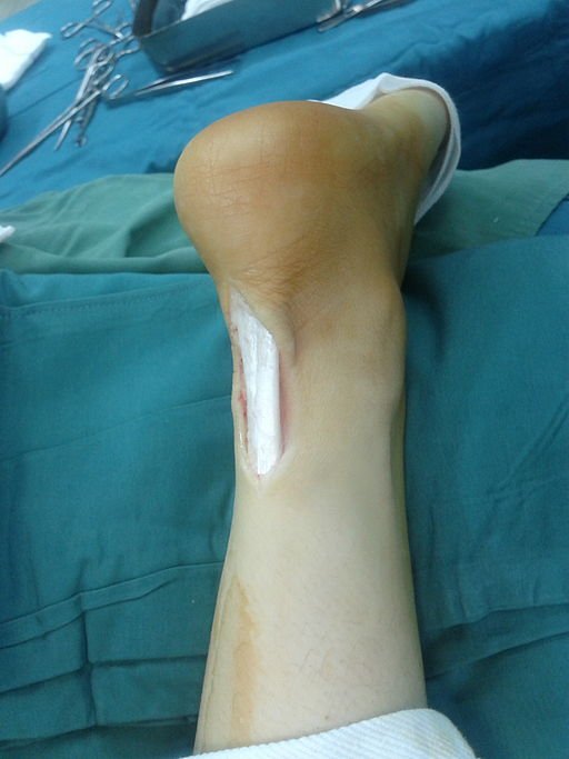
Achilles, Achilles Tendon, and Heel Tendon — Is There a Difference?
This is a very common question, and the answer is simple: there is no difference whatsoever. These are all different names for the same tendon. “Achilles” is the most internationally recognized name, derived from the Greek mythological hero whose only vulnerable point was his heel. In Arabic medical literature, it is also called “watar al-urqub” (the heel tendon), which is the formal anatomical Arabic term. Regardless of which name you search for, you are looking for the same condition.
Causes of Achilles Tendonitis
Achilles tendonitis does not typically result from a single sudden injury. Instead, it develops gradually due to multiple contributing factors:
- Overuse: The most common cause. Sudden increases in training intensity, duration, or frequency — such as rapidly increasing running distance.
- Inappropriate footwear: Worn-out shoes, lack of arch support, or shoes not designed for the specific sport being practiced.
- Gout: Elevated uric acid levels lead to crystal deposits around joints and tendons.
- Tight or shortened calf muscles: Tight gastrocnemius and soleus muscles place enormous, sustained pressure on the Achilles tendon.
- Uneven training surfaces: Uphill running or exercising on uneven terrain increases tendon strain.
- Aging: Blood flow to tendons diminishes with age, and tendons lose some of their elasticity, making them more susceptible to injury.
- Structural abnormalities: Flat feet or a bony prominence at the back of the heel (Haglund’s deformity) causing mechanical friction against the tendon.
Symptoms of Achilles Tendonitis
Symptoms typically develop gradually and may start as mild discomfort before worsening over time if left untreated:
- Morning pain: Severe pain and stiffness at the back of the heel upon taking the first steps in the morning.
- Activity-related pain: Pain begins as mild discomfort at the start of exercise, may disappear during activity, but returns intensely afterward or the following day.
- Tenderness to touch: The tendon becomes very painful when pressed or touched directly.
- Swelling: Visible swelling or thickening of the tendon tissue, noticeable when compared with the opposite foot.
- Pain on stairs: Pain when climbing stairs or standing on tiptoes.
Complications of Achilles Tendonitis
“Toughing out” the pain or “learning to live with it” in Achilles tendonitis is a dangerous decision. Ignoring symptoms can lead to severe complications:
- Chronic tendinopathy: Acute inflammation transforms into chronic degeneration of tendon tissue. The tendon becomes weak, permanently swollen, and painful with even the simplest daily activities.
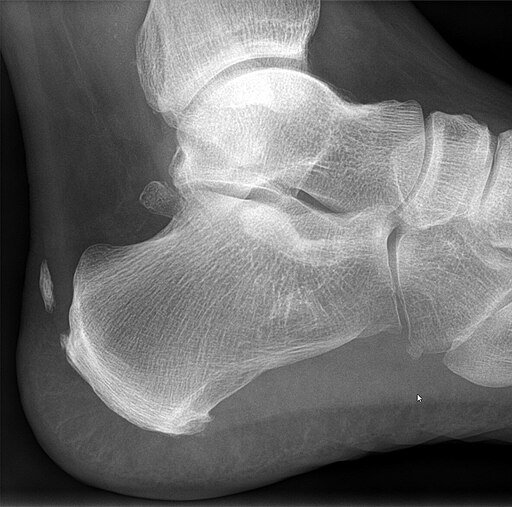
- Bone spurs: The body may attempt to “repair” chronic inflammation by forming small bony deposits at the tendon’s attachment to the heel, increasing pain and friction.
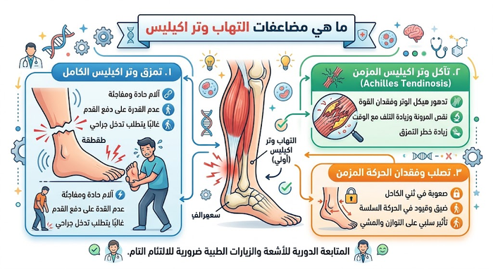
- Complete tendon rupture — the most dangerous complication: An inflamed, weakened tendon is far more susceptible to rupture. The tear may occur suddenly during a simple movement or jump, and the patient feels as though they received a “hard blow” to the heel, losing the ability to walk. This is an emergency that typically requires surgical repair.
Best Treatment for Achilles Tendonitis
The best treatment is one that begins early and addresses the root cause — not just the symptoms.
Phase One: Conservative Treatment (The Foundation)
- Rest: The most critical step. Immediately stop the pain-causing activity (such as running) and substitute with low-impact exercises that do not stress the tendon (swimming or cycling).
- RICE Protocol: Rest, Ice (15–20 minutes several times daily), Compression (light bandage or ankle brace to limit movement and swelling), and Elevation (raise the affected foot above heart level).
- Physical therapy: Treatment is incomplete without it. The physiotherapist focuses on eccentric exercises — scientifically proven to rebuild and strengthen tendon tissue — and stretching exercises to lengthen tight calf muscles and relieve tendon pressure.
- Medication: Short-term use of NSAIDs (such as ibuprofen or diclofenac — oral or topical) for temporary pain and inflammation relief; these do not address the underlying problem.
Phase Two: Advanced Treatments (If Conservative Measures Fail)
If pain persists beyond 3–6 months, your physician may recommend:
- Platelet-rich plasma (PRP) injection: One of the newest and safest approaches. A sample of the patient’s own blood is drawn, the platelet-rich plasma containing growth factors is separated, and it is injected directly into the damaged tendon to stimulate natural healing.
- Extracorporeal shockwave therapy (ESWT): High-energy sound waves are used to stimulate blood flow and promote healing within the tendon.
Phase Three: Surgical Intervention
Surgery is reserved as a last resort for chronic cases unresponsive to all other treatments, or when significant partial tears are present. Surgical options include debridement (cleaning the tendon and removing degenerated tissue) or tendon grafting when damage is extensive.
Can I Exercise with Achilles Tendonitis?
The short answer: do not exercise through pain. The golden rule is: “Do not train with Achilles tendon pain.” Running or jumping on an inflamed tendon worsens the condition, delays healing, and increases the risk of rupture.
- What is permitted: Low-impact sports that do not stress the tendon, such as swimming or cycling (with proper seat positioning), provided they cause no pain.
- When to return to running: The return must be very gradual, only after pain has completely resolved, and under the supervision of the treating physician or physiotherapist.

Conclusion: Do Not Ignore the Pain
Achilles tendonitis is a frustrating injury, but it is entirely treatable when managed correctly and early. Listening to your body and stopping when you feel pain is the first and most important step. Do not let heel pain stop you from moving. Accurate diagnosis and early treatment are the keys to safely returning to your normal life and athletic activities.
Frequently Asked Questions
What is Achilles tendonitis and what is the difference between insertional and non-insertional types?
The Achilles tendon is the thickest and most powerful tendon in the human body, joining the gastrocnemius and soleus muscles to the calcaneus. The condition is clinically categorized by anatomical location:
- Non-Insertional Achilles Tendinopathy: Localized in the mid-substance 2 to 6 cm proximal to the calcaneal insertion. This zone is a vascular “watershed area” with naturally low baseline perfusion, making it susceptible to micro-trauma, tendinosis, and collagen matrix breakdown in runners and jumping athletes.
- Insertional Achilles Tendinopathy: Involves the direct osteotendinous junction on the posterosuperior aspect of the calcaneus. It is often accompanied by retrocalcaneal bursitis, enthesophytes (traction bone spurs), and Haglund’s deformity (a prominent posterosuperior calcaneal tuberosity).
What are the primary causes and biomechanical risk factors for Achilles tendonitis?
Achilles tendinopathy is an overuse disorder driven by cumulative mechanical loading exceeding tissue repair capacity:
- Training Errors: Rapid increases in running volume, abrupt introduction of hill work, or aggressive plyometric routines without adequate recovery.
- Muscle & Joint Kinematics: Gastrocnemius-soleus contracture and restricted ankle dorsiflexion, which impose elevated tensile strain during terminal stance.
- Foot Morphology: Excessive foot pronation (causing an asymmetric “whipping” torque through the tendon) or rigid cavus foot structures with poor shock attenuation.
- Medication Vulnerability: Systemic use of fluoroquinolone antibiotics (such as ciprofloxacin or levofloxacin) within the preceding 6 months, which destabilizes extracellular matrix integrity.
What classic symptoms indicate Achilles tendonitis rather than a muscle strain?
Patients typically present with progressive, load-dependent localized complaints:
- Morning Stiffness & “First-Step” Pain: Marked tightness and pain on the initial steps after waking, which temporarily eases as local tissue temperature and mobility increase.
- Warm-Up Phenomenon: Pain that flares up at the initiation of a run, blunts during the workout, and re-emerges with higher intensity several hours post-exercise.
- Focal Thickening: Palpable swelling, localized heat, or a discrete nodule along the tendon body that moves synchronously with plantarflexion and dorsiflexion.
- Functional Impairment: Difficulty executing a single-leg heel raise or pain during push-off while climbing stairs.
How do orthopedic doctors distinguish Achilles tendonitis from an acute tendon rupture?
Accurate differentiation between tendinopathy, partial tears, and complete rupture is essential:
- The Thompson Test: With the patient prone and knee bent at 90°, the clinician squeezes the calf muscle. If the foot fails to passively plantarflex, an acute full-thickness rupture is confirmed.
- Musculoskeletal Ultrasound (MSK US): Provides dynamic, in-office visualization of tendon diameter, fiber discontinuity, hypoechoic tissue pockets, and Doppler flow (neovascularization) in chronic tendinosis.
- Magnetic Resonance Imaging (MRI): Yields cross-sectional details regarding longitudinal intrasubstance splits, deep retrocalcaneal bursal effusion, and surrounding paratenon inflammation.
Why are direct corticosteroid injections strictly contraindicated for Achilles tendonitis?
Corticosteroid injections into or immediately adjacent to the Achilles tendon are contraindicated:
While steroids offer short-term anti-inflammatory relief, their histological impact on weight-bearing tendons is destructive. High local steroid concentrations suppress tenocyte viability, downregulate type I collagen synthesis, and alter mechanical load tolerance.
Clinical registries show a significant correlation between local corticosteroid infiltration and subsequent catastrophic, non-traumatic tendon rupture during normal daily ambulation. Consequently, modern guidelines endorse biological, physical, and mechanical modalities over local steroid injections.
What is the evidence-based role of eccentric loading (Alfredson Protocol) in tendon recovery?
Eccentric strength training is the foundation of non-surgical Achilles rehabilitation:
- Mechanics (Alfredson Protocol): The patient raises onto the balls of both feet on the edge of a step, lifts the unaffected leg, and slowly lowers the symptomatic heel beneath step level over a 3-second cadence (performing 3 sets of 15 repetitions, twice daily, with both straight and bent knee positions).
- Biological Remodeling: Controlled eccentric elongation produces mechanotransduction signals that stimulate tenocytes to lay down organized collagen fibers, while progressively downregulating pain-generating neovessels and accompanying nerve sprouting.
What advanced non-surgical and biological treatments exist for chronic Achilles tendinopathy?
For refractory tendinopathy resisting first-line rehabilitation, specialized adjunctive modalities are utilized:
- Extracorporeal Shockwave Therapy (ESWT): Acoustic energy waves applied to the chronic nodule to stimulate neo-angiogenesis, disrupt calcific deposits, and downregulate sensory substance P.
- Ultrasound-Guided Platelet-Rich Plasma (PRP): Autologous centrifuged platelets injected precisely into hypoechoic degenerative pockets to deliver concentrated growth factors (such as PDGF, TGF-β) that support cellular healing.
- High-Volume Image-Guided Injections (HVIGI): Saline-based mechanical stripping under ultrasound to shear aberrant neovascular and neural networks out of the pre-Achilles fat pad.
- Orthotic Heel Lifts: 10 to 12 mm viscoelastic heel wedges placed inside running shoes to temporarily unload tensile stress during the rehabilitation period.
When is surgical intervention indicated for Achilles tendonitis?
Surgery is reserved for chronic cases that fail non-operative treatment:
- Clinical Threshold: Persisting symptoms that limit ambulation despite at least 6 months of continuous, compliant eccentric rehabilitation and biological therapies.
- Non-Insertional Debridement: Open or minimally invasive endoscopic excision of fibrotic, mucoid degenerative tissue, followed by longitudinal paratenon release and tubular re-suturing.
- Insertional Reconstruction: Resection of retrocalcaneal bursal tissue, osteotomy of the posterosuperior calcaneal spur (Haglund’s deformity), and tendon re-anchoring using suture anchors.
- FHL Tendon Transfer: If degenerative debridement requires resecting more than 50% of the tendon body, the flexor hallucis longus (FHL) tendon is mobilized to augment plantaris strength and vascularity.
How long does it take for Achilles tendonitis to heal completely?
Tendon recovery timelines reflect the low vascularity and slow baseline turnover of collagen structures:
- Acute Tendonitis (Early Inflammatory): Typically stabilizes within 4 to 8 weeks through load regulation, cryotherapy, and calf flexibility exercises.
- Chronic Tendinosis (Degenerative Matrix): Requires 3 to 6 months of progressive eccentric loading to remodel the structural extracellular matrix.
- Post-Surgical Recovery: Involves a period in a protective CAM walking boot with gradual heel-wedge reduction, followed by physical therapy. High-demand running, plyometrics, and competitive sports generally resume between 6 and 9 months post-op.
How can athletes and runners prevent recurrent Achilles tendon injuries?
Proactive load monitoring and mechanical preparation protect the tendon from recurrent injury:
- Adhere to the 10% Progression Rule: Avoid expanding weekly running mileage, speed intervals, or elevation gain by more than 10% in a single training week.
- Maintain Eccentric Calf Capacity: Incorporate heavy, slow resistance calf raises and eccentric drops as maintenance work 1 to 2 times weekly year-round.
- Footwear Transitions: Avoid abruptly shifting from traditional training shoes (10–12 mm heel-to-toe drop) to minimalist, zero-drop footwear, which spikes tensile strain on unconditioned tendons.
- Cross-Training: Integrate non-impact aerobic conditioning (such as swimming, rowing, or cycling) on active recovery days to offload the ankle joint.
References
AAOS OrthoInfo — Achilles Tendinitis (2024).
https://orthoinfo.aaos.org/en/diseases–conditions/achilles-tendinitis/- AAOS OrthoInfo — Achilles Tendon Rupture (Tear) (2024).
https://orthoinfo.aaos.org/en/diseases–conditions/achilles-tendon-rupture-tear - Cleveland Clinic — Achilles Tendinitis: Symptoms & Treatment (2024).
https://my.clevelandclinic.org/health/diseases/21553-achilles-tendinitis - Cleveland Clinic — Achilles Tendon Rupture: What Is It, Symptoms & Treatment (2024).
https://my.clevelandclinic.org/health/diseases/21703-achilles-tendon-rupture - Mayo Clinic — Achilles Tendinitis: Symptoms & Causes (2024).
https://www.mayoclinic.org/diseases-conditions/achilles-tendinitis/symptoms-causes/syc-20369020 - Hospital for Special Surgery (HSS) — Achilles Tendon Pain: Causes, Diagnosis and Treatment (2024).
https://www.hss.edu/health-library/conditions-and-treatments/list/achilles-tendon - BMC Musculoskelet Disord — The efficacy of eccentric exercise in the treatment of Achilles tendinopathy: a systematic review and meta-analysis. (2026). PubMed 42021217.
https://pubmed.ncbi.nlm.nih.gov/42021217/ - Orthopaedic Journal of Sports Medicine — Efficacy of Platelet-Rich Plasma in Treatment of Achilles Tendinopathy: Systematic Review and Meta-Analysis. (2025). PubMed 40161095.
https://pubmed.ncbi.nlm.nih.gov/40161095/
§6 — English Editorial Version