
Quick answer
Rickets is soft bone in a growing child, most often caused by vitamin D or calcium deficiency. It shows as bowed legs, widened wrists, late walking and teething, and beading of the ribs. It is diagnosed with blood tests and wrist or knee X-rays and treated with vitamin D and calcium for at least 12 weeks; most bowing improves without surgery. Daily vitamin D drops from birth prevent it.
✅ Last medically reviewed by Prof. Dr. Ibrahim Shaarawi, Professor of Orthopaedic Surgery, on 5 October 2026
As an orthopaedic surgeon who treats children’s limb deformities, I see children brought in for bowed legs, and rickets explains a share of them. The first step is to separate normal, age-related bowing from pathological bowing; here we focus on rickets and its link to vitamin D deficiency.
What is rickets?
Rickets is defective mineralisation of bone and growth plates in children. Because bone grows quickly, a lack of calcium and phosphate widens the growth plates and softens bone, which bends under the child’s weight. It is the childhood counterpart of osteomalacia.
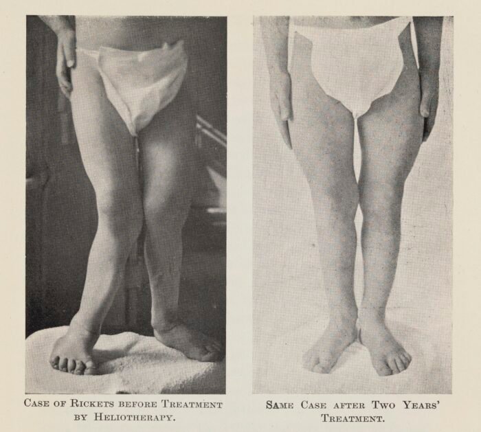
Signs and symptoms of rickets (with pictures)
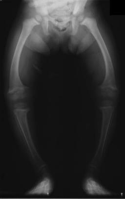
- Bowed legs or knock knees that worsen with walking.
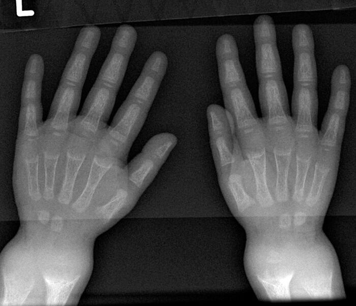
- Widened wrists and ankles from enlarged growth plates.
- Rachitic rosary: bead-like swellings where the ribs meet the cartilage.
- Delayed closure of the soft spot, frontal bossing, and delayed sitting, walking and teething.
- Bone pain, muscle weakness, irritability and poor growth.
- In infants: seizures or tremor from low calcium — an emergency.
Video: which vitamin D dose should I take?
Causes and risk factors
- Exclusive breastfeeding without vitamin D drops, because breast milk is low in vitamin D.
- Maternal vitamin D deficiency during pregnancy and breastfeeding.
- Little sun exposure, darker skin and staying indoors.
- Low dairy and calcium intake after weaning.
- Prematurity, and malabsorption, liver or kidney disease.
- Rare inherited forms (vitamin D-resistant or hypophosphataemic rickets) that do not respond to standard treatment.
Deficiency is common in Egypt: in a study of 572 healthy Egyptian adolescents, more than 94% were vitamin D deficient, especially girls and those with little activity and sun exposure.
Diagnosis: blood tests and X-rays
- Blood tests: 25-hydroxyvitamin D, calcium, phosphate, alkaline phosphatase (usually high) and parathyroid hormone.
- Wrist or knee X-ray: widened growth plates with cupping and fraying of the bone ends.
Key numbers to know
- The Endocrine Society (2024) suggests vitamin D supplements for children aged 1–18 to prevent rickets, for adults over 75, and during pregnancy (Demay et al., Endocrine Society 2024).
- In a study of Egyptian women of childbearing age, 43% were vitamin D deficient and 13% insufficient; only 44% had normal levels (Gerges et al., 2021).
| Treatment | When we use it | Notes |
|---|---|---|
| Treatment doses of vitamin D with calcium | Osteomalacia or rickets due to vitamin D deficiency | Pain and blood tests improve over weeks to months |
| Treating the cause | Malabsorption, kidney or liver disease, certain medicines | Essential to prevent recurrence |
| Follow-up blood tests and X-rays | Throughout treatment | Calcium, phosphate, alkaline phosphatase and vitamin D |
| Surgical correction of bowing | Severe bowing that persists after treating the cause (rare) | Most cases improve with medical treatment |
At Prof. Dr. Ibrahim Shaarawi’s clinics in Nasr City, Mohandeseen and New Cairo (5th Settlement) we review your vitamin D, calcium and bone density results and examine the cause of pain or fracture before deciding on the right dose or osteoporosis treatment.
Treatment: doses and duration
Under the global consensus recommendations for nutritional rickets, vitamin D is given for at least 12 weeks with calcium (about 500 mg daily from food or supplements), followed by a maintenance dose:
| Age | Daily treatment dose | Or single large dose | Maintenance after treatment |
|---|---|---|---|
| Under 3 months | 2,000 IU | Not recommended | 400 IU |
| 3–12 months | 2,000 IU | 50,000 IU | 400 IU |
| 1–12 years | 3,000–6,000 IU | 150,000 IU | 600 IU |
| Over 12 years | 6,000 IU | 300,000 IU | 600 IU |
Blood tests and a wrist X-ray are repeated after about 3 months to confirm healing. Drop doses and common products are covered in vitamin D for infants and children.
Do bowed legs from rickets need surgery?
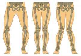
In most children bowing improves gradually after rickets is treated, as bone remodels with growth. Surgery is considered only if a clear deformity persists after healing and adequate follow-up — either guided growth with small plates at the growth plates or a corrective osteotomy in older children.
Prevention
- Vitamin D drops 400 IU daily from the first days of life, especially for breastfed babies.
- Treat maternal deficiency during pregnancy.
- A calcium-rich diet after weaning (milk, yoghurt, cheese).
- Outdoor play and regular physical activity.
Frequently asked questions about rickets in children
What are the symptoms of rickets in babies?
Delayed closure of the soft spot, late sitting and teething, widened wrists, beading of the ribs, irritability and, in severe cases, seizures from low calcium.
Does rickets cause bowed legs?
Yes, rickets is a leading pathological cause of bowed legs, but bowing can be normal until about age two. Persistent or worsening bowing after that needs assessment.
Do bowed legs go away after rickets treatment?
In most children they improve gradually with growth after the deficiency is treated. Surgery is needed only if a clear deformity remains after healing and follow-up.
How long is rickets treated for?
Treatment-dose vitamin D for at least 12 weeks with calcium, then a daily maintenance dose, with blood tests and an X-ray after about 3 months.
How can I prevent rickets?
Give vitamin D drops 400 IU daily from birth, treat maternal deficiency, offer a calcium-rich diet and encourage outdoor play.
To have your bones assessed and your tests reviewed, book a consultation, call +20 155 240 4488 or message us on WhatsApp at +20 155 554 7181.
Medically reviewed by Prof. Dr. Ibrahim Shaarawi, Professor of Orthopaedic Surgery. This article is general medical education and does not replace an examination or the treatment plan your doctor sets for you. See our medical disclaimer.
Related vitamin D and bone health guides
Related guides on rickets and vitamin D:
- Vitamin D deficiency
- Vitamin D injections
- Vitamin D deficiency and bone and joint pain
- Osteomalacia
- Calcium injections for bones
- Vitamin D for infants and children
- The best vitamins for bones and joints
- Osteoporosis
References
- Munns CF, et al. Global Consensus Recommendations on Prevention and Management of Nutritional Rickets. J Clin Endocrinol Metab. 2016;101(2):394-415.
- Uday S, Högler W. Nutritional Rickets and Osteomalacia in the Twenty-first Century: Revised Concepts, Public Health, and Prevention Strategies. Curr Osteoporos Rep. 2017;15(4):293-302.
- Sherief LM, et al. Vitamin D status and healthy Egyptian adolescents: Where do we stand? Medicine (Baltimore). 2021;100(29):e26661.
- Demay MB, et al. Vitamin D for the Prevention of Disease: An Endocrine Society Clinical Practice Guideline. J Clin Endocrinol Metab. 2024;109(8):1907-1947.