
Heel Spur (Plantar Fasciitis) Treatment is one of the most frequently asked questions by patients with plantar fasciitis. This guide covers the best at-home exercises, when to see a doctor, and how to maximise results.
The most important step in treating plantar fasciitis is identifying and stopping the cause. Exercises alone will not produce lasting results if the underlying risk factor continues.
Causes and Risk Factors
- Walking barefoot on hard surfaces
- Wearing high heels
- Prolonged standing
- Excess body weight
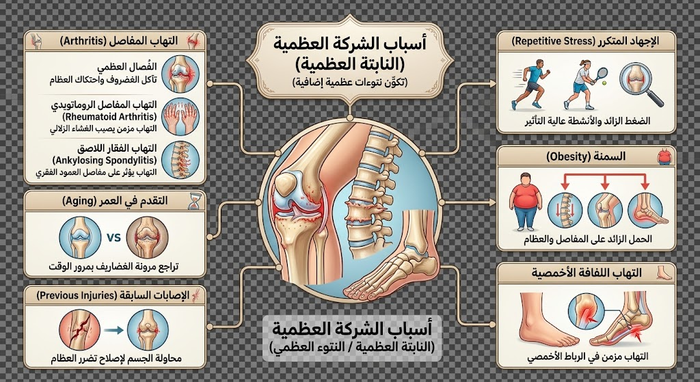
Risk Factor Detail and Modifiability
| Risk Factor | Mechanism | Modifiable? | First Corrective Step |
|---|---|---|---|
| Barefoot walking on hard floors | Direct load without shock absorption | Immediately | Wear supportive footwear at all times |
| High heels (> 5 cm) | Chronically shortens calf muscle and Achilles | Immediately | Transition gradually to 2–3 cm heel |
| Prolonged standing (> 6 hr/day) | Repetitive plantar fascia stress | Yes | Rest breaks + cushioned anti-fatigue mat |
| Excess weight (BMI > 25) | Each extra kg adds 3–4 kg of heel pressure | Gradually | Weight reduction + low-impact exercise (swimming, cycling) |
| Flat foot or high arch | Abnormal load distribution across fascia | Partially | Custom orthotic insole |
| Running on hard surfaces or sudden training increase | Repetitive micro-tears at fascia origin | Yes | Gradual load progression + running shoes |
| Age over 40 | Reduced fascia and tendon elasticity | No | Daily stretching as prevention |
| * Based on AAOS, Mayo Clinic, and APMA guidelines 2024. Addressing modifiable causes before starting exercises significantly improves outcomes. | |||
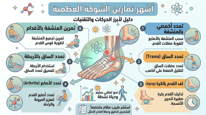
The 6 Best Home Exercises for Plantar Fasciitis
1. Towel Toe Curls
- Place a small towel flat on the floor. Using only the toes of the affected foot, scrunch and pull the towel towards you, then release.
- Repeat 10 times per session, 5 sessions per day.
2. Seated Toe Stretch
- Sit and cross the affected foot over your other leg. Hold your toes and pull them upward as far as comfortable to stretch the plantar fascia and calf. With the other hand, massage the arch of the foot.
- Hold 10 seconds per stretch; continue for 5 minutes total, 5 sessions per day.
3. Standing Calf Stretch
- Stand facing a wall for support. Place the affected leg behind and the other in front, knee slightly bent. Keep the rear heel flat on the floor and lean forward until you feel a stretch in the calf.
- Hold 45 seconds, 2–3 repetitions, 5 sessions per day.
4. Towel Stretch (Best Done Before Getting Out of Bed)
- Sit with the affected leg extended. Loop a towel around the foot and gently pull toward you until you feel a calf and arch stretch.
- Hold 45 seconds, 10 repetitions, 5 sessions per day.
5. Stair Heel Drop
- Stand on a step with the unaffected foot fully supported. Place only the front of the affected foot on the step edge and slowly lower the heel below step level until a calf stretch is felt.
- Hold 45 seconds, 2–3 repetitions, 4–6 sessions per day.
6. Ice Bottle Massage
- While seated, roll the affected foot over a frozen water bottle back and forth.
- Continue 3–5 minutes, 5 sessions per day (best after other exercises or at end of day).
Daily Exercise Protocol — Dose and Timing
| Exercise | Hold / Duration | Reps per Session | Sessions / Day | Best Timing | Target Structure |
|---|---|---|---|---|---|
| Towel toe curls | Movement | 10 reps | 5 | Any time — seated | Intrinsic foot muscles |
| Seated toe stretch | 10 sec | 5 min continuous | 5 | Morning before first step — most important | Plantar fascia + Achilles |
| Standing calf stretch | 45 sec | 2–3 | 5 | Spread throughout day | Gastrocnemius |
| Towel stretch | 45 sec | 10 | 5 | Morning before first step — very important | Plantar fascia + Soleus |
| Stair heel drop | 45 sec | 2–3 | 4–6 | After warm-up only — not cold | Gastrocnemius + Soleus |
| Ice bottle massage | 3–5 min | 1 | 5 | After exercises or end of day | Inflammation and local pain relief |
| * Morning stretching before the first step is most critical — the plantar fascia contracts overnight and needs gradual lengthening. Results typically emerge after 6–8 weeks of daily adherence. Source: Washington University Orthopaedics, AAOS, DiGiovanni et al. JBJS 2006. | |||||
Treatment Options — From Home to Clinical Intervention
| Treatment Option | Setting | Pain Relief Rate | Time to Results | Best For | Limitation |
|---|---|---|---|---|---|
| Home stretching programme | Home | 70–80% with adherence | 6–8 weeks | New onset (< 6 months) — mild to moderate | Requires strict daily discipline |
| Custom orthotic insole | Podiatrist / orthopaedic | 60–75% | 2–4 weeks | Flat foot or high arch | Cost + requires prescription |
| Oral NSAIDs | Pharmacy | Temporary only — not curative | Days | Acute pain control | GI side effects — does not treat cause |
| Corticosteroid injection | Specialist clinic | 70–90% temporary | 1 week | Failed home treatment after 6 weeks | Max 2–3 injections — tendon rupture risk |
| Extracorporeal shockwave therapy (ESWT) | Specialist clinic | 60–80% in chronic cases | 4–12 weeks | Chronic (> 6 months) — failed conservative | Not universally available |
| Surgical fascia release | Hospital | 75–90% in selected cases | 2–4 months | Failed all conservative care > 12 months | Last resort — anaesthesia + rare tendon injury |
| * Success rates from AAOS, Mayo Clinic, and Cochrane Reviews. Over 90% of plantar fasciitis patients recover with conservative care within 12 months without surgery. | |||||
When to Stop Home Exercises and See a Doctor
Stop exercises and seek evaluation if any of these are present:
- No improvement after 6–8 weeks of daily exercises
- Pain worsening with exercise (possible partial fascial tear)
- Tingling or numbness in the foot or ankle (nerve involvement)
- Visible swelling, redness, or warmth (infection or inflammatory arthritis)
- Pain extending up the leg, not just the heel
- Diabetic patient or immunocompromised — any heel pain warrants immediate evaluation
Frequently Asked Questions
What is the relationship between a heel spur and plantar fasciitis?
A heel spur and plantar fasciitis are closely linked clinical entities with distinct pathological definitions:
- Plantar Fasciitis: The primary source of symptoms; an inflammatory and degenerative micro-tearing of the plantar aponeurosis originating at the medial calcaneal tuberosity.
- Heel Spur (Calcaneal Spur): A reactive osteophytic traction hook that forms when chronic fascial tension pulls on the calcaneal periosteum, prompting adaptive calcium deposition over months or years.
- Clinical Reality: Up to 10% to 15% of the general population have radiographic heel spurs without any pain, and resolving fascial inflammation eliminates symptoms completely without removing the spur.
Why is heel spur pain sharpest during the first steps in the morning?
The hallmark “first-step pain” (post-static dyskinesia) is governed by nighttime tissue changes:
- Nocturnal Shortening: During sleep, the ankle naturally rests in passive plantarflexion, causing the inflamed plantar fascia and calf musculature to relax and shorten during micro-repair.
- Sudden Tensile Stress: Taking the first steps out of bed abruptly loads body weight across the shortened fascia, re-tearing delicate collagen bridges and firing high-intensity nociceptive pain signals.
- Dynamic Warm-Up: Continued walking warms up the tissue and gradually elongates the fibers, diminishing the sharp stabbing pain into a dull background ache before it potentially returns by evening.
What are the primary causes and biomechanical risk factors for heel spurs?
Heel spur pathogenesis involves chronic biomechanical loading and soft-tissue tightness:
- Elevated BMI & Sudden Weight Gain: Increases vertical ground reaction forces and accelerates repetitive micro-trauma across the longitudinal arch.
- Occupational Demands: Prolonged weight-bearing or standing on rigid, concrete industrial flooring.
- Foot Morphology Abnormalities: Severe flat feet (overpronation causing excessive fascial stretch) or rigid high arches (poor shock absorption).
- Triceps Surae Tightness: Shortened gastrocnemius, soleus, or Achilles tendon fibers that restrict ankle dorsiflexion and place compensatory tension on the plantar fascia.
- Improper Footwear: Wearing unsupportive, flat shoes or walking barefoot on hard tile surfaces.
How is plantar fasciitis with a heel spur diagnosed clinically and radiologically?
A comprehensive diagnosis combines physical evaluation with targeted imaging:
- Palpation: Focal, exquisite point tenderness directly over the anteromedial aspect of the inferior heel.
- Windlass Test: Passive hyperextension of the great toe while the patient stands or reclines, which stretches the aponeurosis and reproduces typical heel discomfort.
- Weight-Bearing X-Rays: Plain radiographs confirm the presence, size, and orientation of the calcaneal spur while ruling out stress fractures and systemic spondyloarthropathies.
- High-Resolution Ultrasound (MSK US): Directly visualizes fascial architecture; a fascial thickness exceeding 4.0 mm (normal is 2.5 to 3.5 mm) with hypoechoic thickening and loss of fibrillar pattern confirms active disease.
What are the most effective daily stretching exercises for plantar fasciitis recovery?
Consistent mechanical stretching is the most critical component of non-surgical recovery:
- Frozen Water Bottle Roll: Roll the arch over a frozen plastic bottle for 10 to 15 minutes twice daily to combine mechanical tissue mobilization with anti-inflammatory cryotherapy.
- Wall-Assisted Gastrocnemius Stretch: Step the affected leg back, keep the knee straight with the heel flat on the floor, and lean forward until a deep pull is felt in the upper calf; hold for 30 seconds across 3 repetitions.
- Pre-Step Morning Towel Stretch: Loop a folded towel or strap beneath the metatarsal heads before getting out of bed, pulling the forefoot toward the chest to pre-stretch the plantar fascia prior to weight-bearing.
How does Extracorporeal Shockwave Therapy (ESWT) treat chronic heel spurs?
ESWT is an advanced non-invasive intervention for chronic, refractory cases:
- Mechanism of Action: High-energy acoustic sound waves are delivered directly into the degenerative fascial insertion, inducing controlled biological microtrauma.
- Biological Cascade: Triggers release of angiogenic growth factors (e.g., VEGF), induces local neovascularization, breaks up micro-calcifications, and accelerates structural collagen synthesis.
- Pain Modulation: Depletes Substance P and hyperstimulates local nociceptors, providing pain relief with success rates reaching 75% to 80% across 3 to 5 weekly sessions.
What is the difference between corticosteroid injections and PRP for heel spurs?
Both injectable options are utilized in clinical practice under distinct therapeutic rationales:
- Corticosteroid Injections:
• Benefits: Powerful, immediate suppression of acute pain and synovial inflammation within 48 hours.
• Drawbacks: Temporary symptomatic relief (lasting weeks to months); carries risks of permanent subcalcaneal fat pad atrophy, skin hypopigmentation, and acute plantar fascia rupture if repeated. - Platelet-Rich Plasma (PRP) Injections:
• Benefits: Autologous concentrated platelets containing growth factors (PDGF, TGF-β) that stimulate genuine biological collagen remodeling and tissue regeneration.
• Drawbacks: Takes 3 to 6 weeks to show peak clinical benefit, but provides permanent repair without tissue weakening.
What shoe features and custom orthotics provide the best relief for heel spurs?
Mechanical offloading of the calcaneal insertion requires specific footwear parameters:
- Footwear Architecture: Running or walking shoes with thick, responsive EVA or polyurethane midsoles, a rigid external heel counter to stabilize the rearfoot, and a mild heel-to-toe drop (8 to 12 mm) to reduce Achilles tension.
- Custom Orthotics & Inserts: Semi-rigid arch supports crafted to control excessive pronation and support the medial longitudinal arch.
- Silicone Heel Cushions: Specialized medical heel cups with a central hollowed-out aperture (“horseshoe” or teardrop recess) that suspends the spur in air, preventing direct ground contact pressure.
Do night splints help alleviate morning heel pain?
Yes, night splints are an evidence-based tool for addressing morning post-static dyskinesia:
- Biomechanics: A rigid or dorsal night splint maintains the foot and ankle in a controlled 90-degree neutral position with slight extension of the toes.
- Prevention of Contracture: By maintaining a gentle stretch overnight, the plantar fascia heals in an elongated state rather than shortening, substantially minimizing the tearing sensation experienced during morning ambulation.
- Compliance: Typically worn consistently for 4 to 8 weeks until morning pain subsides.
When is surgical intervention necessary for a heel spur or plantar fasciitis?
Surgery is reserved as an end-stage option for refractory cases:
- Indications: Considered only when severe, recalcitrant heel pain persists after 6 to 12 months of exhaustive conservative therapy (stretching, orthotics, ESWT, and biologics).
- Surgical Technique: Performed via minimally invasive or endoscopic approaches (Endoscopic Plantar Fasciotomy – EPF). The surgeon performs a partial release of the medial 30% to 50% of the fascial band to alleviate tension while preserving lateral column stability.
- Spur Resection: The bony spur is removed only if it is exceptionally prominent or impinging directly on the first branch of the lateral plantar nerve (Baxter’s nerve).
References
- AAOS OrthoInfo. Plantar Fasciitis and Bone Spurs. 2023. Available at: orthoinfo.aaos.org
- Mayo Clinic Staff. Plantar fasciitis – Diagnosis and treatment. 2023. Available at: mayoclinic.org
- Cleveland Clinic. Plantar Fasciitis: Causes, Symptoms, Diagnosis & Treatment. 2023. Available at: my.clevelandclinic.org
- Washington University Orthopaedics. Plantar Fasciitis Exercises. Available at: ortho.wustl.edu
- Hospital for Special Surgery (HSS). Plantar Fasciitis: Symptoms, Causes and Treatment. 2023. Available at: hss.edu
- Medscape. Plantar Fasciitis Treatment & Management. 2024. Available at: emedicine.medscape.com
- Landorf KB, Menz HB. Plantar heel pain and fasciitis. BMJ Clinical Evidence. 2008;2008:1111. Available at: ncbi.nlm.nih.gov
- DiGiovanni BF, et al. Plantar fascia-specific stretching exercise improves outcomes in patients with chronic plantar fasciitis. Journal of Bone and Joint Surgery. 2006;88(8):1775–1781. DOI: 10.2106/JBJS.E.01281