
Sudden knee pain is a common complaint across every age group. It can follow an injury — a torn ACL, a meniscal tear, a fracture — or arise from an underlying condition such as osteoarthritis, gout, or septic arthritis. Many milder cases respond to self-care, physical therapy and cold packs, but some require imaging, aspiration, injections, or surgery. This guide explains the causes by age, the warning signs that need urgent review, the diagnostic pathway, and the modern treatment options available in Cairo.
Symptoms that often accompany sudden knee pain
- Swelling and stiffness — suggest injury or joint inflammation.
- Redness and warmth on touch — suggest bacterial (septic) infection.
- Weakness or instability — classic for a torn cruciate ligament.
- Clicking or popping sounds — often indicate a meniscal tear.
- Inability to fully straighten the knee — can be caused by a mechanical block from a meniscal fragment or by joint effusion.
When to see a knee specialist
Call your doctor if you cannot bear weight on the knee, if the knee feels unstable or gives way, if there is obvious swelling, if you cannot fully bend or straighten it, if the leg looks deformed, if there is fever alongside redness and swelling, or if the pain followed a definite injury and is severe.
Causes of sudden knee pain
Knee injuries
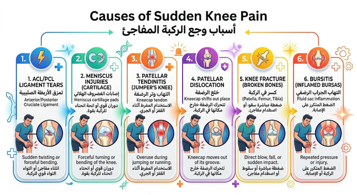
- ACL injury — a tear of the anterior cruciate ligament, common in football, basketball and any sport with rapid direction changes.
- Fractures — of the femur, tibia or patella, from falls or road accidents; in osteoporotic bone, a mis-step alone can cause a fracture.
- Meniscal tear — the meniscus is the shock-absorbing cartilage between femur and tibia. A sudden twist while weight-bearing can tear it.
- Bursitis — inflammation of the small fluid-filled sacs (bursae) that cushion tendons and ligaments as they glide over the joint.
Mechanical problems
- Loose body — a fragment of bone or cartilage floats free in the joint and can lock movement, like a pencil jammed in a door hinge.
- Iliotibial band syndrome — the tough IT band running from the pelvis to the outer knee tightens and rubs against the outer femur, common in distance runners and cyclists.
- Patellar dislocation — the kneecap slips out of its groove, usually laterally. It may reduce spontaneously or remain dislocated.
Osteoarthritis of the knee
The most common form of knee arthritis. Cartilage wears down with age and use, causing pain, stiffness and reduced range.
Rheumatoid arthritis
An autoimmune, chronic condition that can affect most joints including the knees, with a fluctuating course.
Gout
Uric acid crystal deposition, most classically at the big toe but often affecting the knee.
Septic (bacterial) arthritis
A joint infection causing pain, swelling and redness, usually with fever and often without a preceding injury. Septic arthritis can destroy cartilage rapidly and is an orthopedic emergency requiring urgent surgical washout.
Patellofemoral pain syndrome
Pain between the kneecap and the femoral groove, common in athletes, young women, patients with patellar maltracking, and older adults with patellofemoral arthritis.
Causes by age group
Under 30: sports injuries, particularly ACL tears and overuse from repetitive training.
30 to 50: meniscal tears (often needing arthroscopic treatment), tendinopathy, synovial inflammation.
Over 50: osteoarthritis, osteoporotic fractures, and degenerative meniscal changes.
Risk factors
Excess weight increases mechanical load on the joint even during walking or stair use. Poor muscle strength or flexibility leaves the joint less protected. Certain sports (skiing, basketball, running) and physically demanding occupations (construction, farming) raise injury risk. A previous knee injury significantly raises the chance of another.
Diagnosis
Clinical examination checks for swelling, tenderness, warmth, range of motion, and ligament integrity. Imaging is chosen by the suspected pathology: plain X-rays for fractures and degenerative changes, CT for subtle bony injury, ultrasound for real-time soft-tissue assessment, and MRI as the definitive tool for ligament, tendon, cartilage, and meniscal injury. Blood tests and joint aspiration (arthrocentesis) are added when infection or crystal arthritis is suspected.
Treatment
Medications
Directed at the underlying cause: analgesics and NSAIDs for pain and inflammation, urate-lowering therapy for gout, disease-modifying drugs for rheumatoid arthritis.
Physical therapy
Strengthens the muscles that stabilise the knee and corrects movement patterns that overload it. Bracing and heel wedges may offload one side of the joint in unicompartmental arthritis.
Injections
Three main options: corticosteroids for acute inflammatory flares (rapid but time-limited), hyaluronic acid to supplement joint lubrication in moderate osteoarthritis, and platelet-rich plasma (PRP) which delivers growth factors and may promote biological healing in early osteoarthritis. The choice depends on the severity of cartilage loss, the patient’s age and activity level, and comorbidities such as diabetes.
Surgery
- Arthroscopic surgery — through small portals, to remove loose bodies, repair or trim the meniscus, or reconstruct torn ligaments.
- High tibial osteotomy — realigning the leg to shift load away from a diseased compartment, preserving the native joint in younger patients with unicompartmental arthritis and correctable malalignment.
- Partial (unicompartmental) knee replacement — replaces only the affected part of the knee; smaller incision, faster recovery than total replacement.
- Total knee replacement — replaces all worn surfaces of the femur, tibia and patella with metal-and-plastic components. Reserved for advanced multicompartmental disease.
Prevention
Maintain a healthy weight, condition the muscles before demanding activity, use correct technique in sport (a few coaching sessions pay off), build quadriceps and hamstring strength and hip balance, and stretch to keep the surrounding muscles flexible. For chronic knee problems, shift to low-impact activity such as swimming or cycling.
Frequently Asked Questions
What are the most common causes of sudden knee pain?
Sudden, acute-onset knee pain typically stems from sudden structural mechanical failure or rapid-onset intra-articular inflammation:
- Ligament Disruptions: Acute tears of the anterior cruciate ligament (ACL), medial collateral ligament (MCL), or posterior cruciate ligament (PCL) resulting from rotational or hyperextension trauma.
- Meniscal Tears: Sharp shearing or twisting of the knee while flexed and weight-bearing, causing radial, longitudinal, or flap tears.
- Patellar Dislocation / Subluxation: The kneecap slips out of the femoral trochlear groove, frequently tearing the medial patellofemoral ligament (MPFL).
- Crystalline Arthropathies: Acute gout or pseudogout (CPPD) causing severe acute synovitis.
- Septic Arthritis: Bacterial colonization of the joint space presenting as an urgent medical emergency.
What does a loud “popping” sound at the time of knee injury indicate?
An audible or palpable “pop” accompanied by immediate giving-way during sports, sudden direction changes, or awkward landings is a classic clinical indicator:
- ACL Rupture: Responsible for more than 70% of acute traumatic pops in active individuals, followed by sudden instability.
- Patellar Dislocation: Caused by the kneecap tearing through the medial retinaculum as it dislodges laterally.
- Extensor Mechanism Rupture: Complete tearing of the quadriceps tendon or patellar tendon, accompanied by immediate inability to perform an active straight leg raise.
- Peripheral Meniscal Detachment: Avulsion of the vascularized meniscal rim from the joint capsule.
Why does the knee swell rapidly within a few hours of acute pain?
The timing of intra-articular swelling serves as a critical diagnostic indicator:
- Hyperacute Swelling (within 2 to 4 hours): Indicates hemarthrosis (blood inside the joint). This points directly to structural damage of richly vascularized tissues, such as an ACL tear, osteochondral fracture, or peripheral meniscosynovial separation.
- Delayed Swelling (12 to 24 hours later): Represents a reactive synovial effusion caused by the synovium overproducing joint fluid in response to mechanical irritation, such as an avascular central meniscal tear or mild chondral contusion.
What is true mechanical knee locking and why is it an urgent condition?
True mechanical knee locking occurs when the knee cannot be fully straightened (loss of terminal extension) due to a physical obstruction within the joint:
- Bucket-Handle Meniscal Tear: A large longitudinal meniscal fragment flips into the intercondylar notch, wedging between the femoral condyle and tibial plateau.
- Loose Bodies: Detached pieces of cartilage or bone floating free in the joint fluid that migrate between articulating surfaces.
- Clinical Caution: Forcing a locked knee straight can crush trapped meniscal tissue, converting a repairable tear into an irreparable one and stripping healthy articular cartilage. Urgent orthopedic evaluation is necessary.
How do doctors differentiate between traumatic knee pain and an acute gout flare?
Clinical history and physical examination differentiate crystalline arthritis from mechanical joint injuries:
- Traumatic Injuries: Preceded by a clear mechanical event (collision, fall, twist). Pain is aggravated by weight-bearing and movement, while systemic vitals and cutaneous skin temperature remain normal.
- Acute Gout / Pseudogout Flare: Strikes abruptly without preceding trauma, often waking the patient in the early morning hours. The knee becomes erythematous (deep red/purple), markedly warm to the touch, severely swollen, and hypersensitive to light contact (such as a bedsheet). Joint aspiration (arthrocentesis) confirms the presence of negatively birefringent monosodium urate crystals.
What immediate first-aid protocol should be followed for sudden knee pain?
Initiate the standard PRICE protocol immediately during the first 48 hours to minimize secondary tissue damage:
- Protect & Rest: Immobilize the joint, discontinue physical activity, and use crutches to avoid full weight-bearing on the affected limb.
- Ice: Apply a cold pack or crushed ice wrapped in a damp towel for 15 to 20 minutes every 2 to 3 hours to induce vasoconstriction and limit bleeding.
- Compress: Wrap the knee evenly with a flexible elastic bandage, ensuring snug support without impeding peripheral arterial blood flow to the foot.
- Elevate: Keep the injured leg elevated above heart level when resting to facilitate venous return and reduce interstitial effusion.
What red-flag symptoms require immediate emergency room evaluation?
Seek immediate emergency evaluation if sudden knee pain is accompanied by any of these warning signs:
- Complete inability to support body weight or an unstable knee that gives out under load.
- Obvious anatomical deformity, limb misalignment, or lateral dislocation of the kneecap.
- Coldness, pale or bluish discoloration in the calf or foot, or absent peripheral foot pulses (indicating popliteal artery or vascular compromise).
- Numbness, tingling, or progressive foot drop (indicating peroneal or tibial nerve injury).
- High fever, chills, and malaise accompanying a red, tense, and hot knee joint (red flags for septic arthritis).
How is the exact cause of sudden knee pain diagnosed?
Orthopedic specialists follow a structured, multi-step diagnostic pathway:
- Physical Examination: Provocative ligament stability testing (Lachman test, anterior/posterior drawer, pivot-shift, and varus/valgus stress) alongside meniscal signs (McMurray and joint-line tenderness).
- Plain Radiographs (X-rays): Initial imaging to rule out acute fractures, avulsion injuries (e.g., Segond fracture), patellar maltracking, and radio-opaque loose bodies.
- Magnetic Resonance Imaging (MRI): The gold-standard modality for visualizing soft-tissue disruptions, classifying meniscal tear patterns, assessing cruciate and collateral ligament integrity, and detecting subchondral bone bruises.
- Diagnostic Joint Aspiration: Analysis of synovial fluid for leukocyte counts, Gram stain, culture, and crystal analysis when septic or crystalline arthritis is suspected.
When does sudden knee pain require urgent arthroscopic knee surgery?
Minimally invasive knee arthroscopy is indicated in several acute clinical scenarios:
- Locked Bucket-Handle Meniscal Tears: Urgent arthroscopic reduction and meniscal repair (suturing) to preserve the functional cushioning of the meniscus.
- Mechanical Loose Bodies: Removal of detached fragments blocking motion to prevent articular surface destruction.
- Complete ACL Tears: Arthroscopic reconstruction utilizing autografts (hamstring, bone-patellar tendon-bone, or quadriceps tendon) to restore knee kinematics and rotatory stability in active patients.
- Septic Arthritis: Emergent arthroscopic joint lavage, synovectomy, and extensive debridement to clear purulent exudates and prevent irreversible cartilage matrix breakdown.
Can continuing to walk on an acutely injured knee cause permanent damage?
Yes, walking through acute knee pain carries significant risks:
When an internal stabilizing structure—such as an ACL or collateral ligament—is compromised, the knee experiences abnormal subluxation forces during gait. Continued loading can convert a stable, repairable meniscal tear into an irregular, complex maceration, shear protective hyaline cartilage off the femoral condyles, and accelerate early post-traumatic osteoarthritis. Use crutches and keep weight off the joint until an accurate clinical and radiological diagnosis is confirmed.
References
- Mayo Clinic. Knee pain — Symptoms and causes. 2026. mayoclinic.org
- American Academy of Orthopaedic Surgeons – OrthoInfo. Common Knee Injuries. 2026. orthoinfo.aaos.org
- Cleveland Clinic. Knee Pain: Causes, Types, Symptoms & Diagnosis، 2024. my.clevelandclinic.org
- Hospital for Special Surgery (HSS). Knee Pain: Causes and Treatment. 2026. hss.edu
- NICE Clinical Knowledge Summaries. Knee pain — assessment. 2023. cks.nice.org.uk
- Kolasinski SL, Neogi T, Hochberg MC, et al. 2019 American College of Rheumatology / Arthritis Foundation Guideline for the Management of Osteoarthritis of the Hand, Hip, and Knee. Arthritis Care & Research،. acrjournals.onlinelibrary.wiley.com
- Bannuru RR, Osani MC, Vaysbrot EE, et al. OARSI guidelines for the non-surgical management of knee, hip, and polyarticular osteoarthritis. Osteoarthritis and Cartilage،؛. oarsijournal.com
- Medscape Reference. Knee Pain — Overview, Anatomy, Clinical Presentation. 2024. emedicine.medscape.com