
Many patients ask about the appearance of a ganglion cyst of the foot and ankle — one of the most recognised problems of the foot and ankle joint. This guide covers everything you need to know.
What Is a Ganglion Cyst of the Foot?
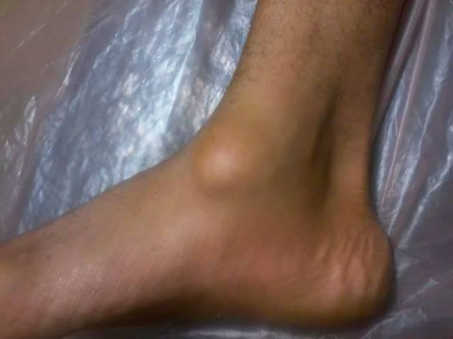
A ganglion cyst is a benign swelling that usually appears adjacent to tendons or joints, particularly in the foot, ankle, wrist, or hand. It takes a round or oval form filled with clear, jelly-like synovial fluid. A ganglion cyst is not cancerous and does not transform into cancer, but it can cause pain or nerve compression in some cases.
Anatomical Locations in the Foot and Ankle
| Location | Typical Appearance | Common Size | Most Common Symptoms | Nerve/Vessel Risk | Evaluation Priority |
|---|---|---|---|---|---|
| Dorsum of foot (top) | Visible round lump | 0.5–3 cm | Shoe conflict — mild pain | Moderate | Watchful waiting or elective |
| Plantar (sole) | Relatively firm — hidden under skin | 0.5–2 cm | Pain on walking — pebble sensation | High — near plantar nerves | Urgent — MRI required |
| Ankle (anterior) | Oval swelling beside the joint | 1–4 cm | Ankle stiffness — pain on movement | Moderate | Assess within one week |
| Posterior ankle (behind heel) | Swelling behind heel — may resemble Baker’s cyst | 1–5 cm | Pain walking and on stairs | High — near posterior tibial nerve | Urgent evaluation |
| Around foot tendons | Elongated mass along tendon course | 1–6 cm | Pain and weakness on toe movement | Moderate | Assess within one week |
| * Plantar and posterior ankle cysts require MRI before any intervention due to proximity to major nerves and vessels. Source: AAOS, Mayo Clinic, HSS. | |||||
Symptoms
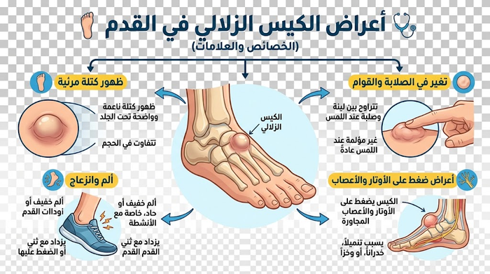
The cyst can be as small as a pea and may change size with activity. It is usually painless, but if it presses on a nearby nerve it may cause pain, tingling, numbness, muscle weakness, and sometimes limited joint movement.
Symptom Classification by Severity
| Symptom | Severity | Impact on Daily Life | Recommended Action |
|---|---|---|---|
| Small cyst < 1 cm — painless — no functional limitation | Mild | Cosmetic concern only | Periodic monitoring every 6 months |
| Shoe conflict — mild pressure pain | Mild–moderate | Footwear restriction | Change footwear + compressive dressing + monitoring |
| Pain during prolonged walking or sport | Moderate | Limits walking and exercise | Assess within one week — aspiration or splinting |
| Tingling or pins and needles in toes | Moderate–severe | Functional impairment | Urgent evaluation — MRI |
| Foot muscle weakness or gait change | Severe | Clear motor disability | Immediate evaluation — nerve compression may require surgery |
| Redness + warmth + severe constant pain | Emergency | Complete impairment — infection risk | Emergency within 24 hours |
| * Any tingling or muscle weakness from a foot ganglion cyst warrants urgent evaluation — chronic nerve compression can cause permanent nerve damage. | |||
Causes
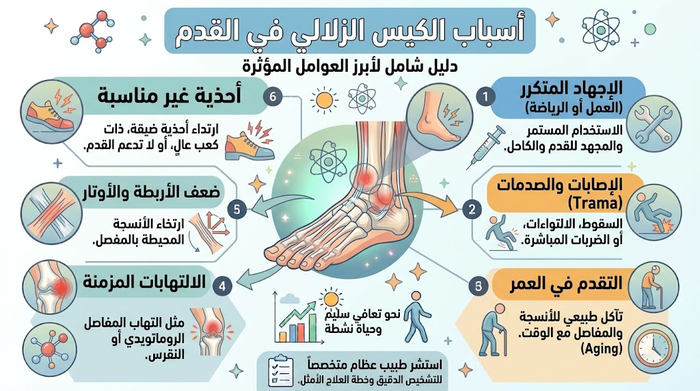
- More common in women, especially aged 20–40.
- Incidence is higher in people with joint osteoarthritis.
- Previous joint or tendon injuries increase the likelihood of occurrence.
Is a Foot Ganglion Cyst Dangerous?
Generally it is not dangerous, but the patient must be examined to confirm the swelling is truly a ganglion cyst — because certain soft-tissue sarcomas can present identically to a ganglion cyst in the hand or foot.
🔴 Important Warning: Soft tissue sarcoma can appear clinically identical to a ganglion cyst in the foot. Any lump that is firm, non-mobile, or growing rapidly must be treated as malignant until MRI and biopsy confirm otherwise.
Diagnosis
Diagnosis is made by clinical examination — the doctor presses on the cyst to assess tenderness, and sometimes shines a light to check content translucency. In some cases imaging is ordered (X-ray, ultrasound, or MRI) to confirm the diagnosis and exclude other causes such as malignant tumours or joint inflammation. A specialist orthopaedic surgeon should always be consulted when any foot swelling is present.
Differential Diagnosis — Foot and Ankle Lumps
| Lump Type | Texture | Changes with Rest | Transillumination | Diagnostic Tool | Malignant Potential |
|---|---|---|---|---|---|
| Ganglion cyst | Soft — fluid-filled | Decreases with rest | Positive (translucent) | Clinical ± ultrasound | Zero — fully benign |
| Lipoma | Soft, rubbery | No change | Negative | Ultrasound | Very low |
| Giant cell tumour of tendon sheath (GCTTS) | Firm | No change | Negative | MRI + biopsy | Rare — requires monitoring |
| Morton’s neuroma | No visible lump — pain only | Improves on removing shoe | N/A | Ultrasound | Zero — benign |
| Soft tissue sarcoma | Firm, fixed | No change | Negative | MRI + mandatory biopsy | Yes — urgent surgery |
| Bursitis | Soft — diffuse | Partial improvement | Variable | Ultrasound | Zero |
| * Key distinguishing feature of a ganglion cyst: size varies with activity/rest + positive transillumination + fluid confirmed on ultrasound. Any firm, non-mobile foot lump requires MRI and biopsy before any intervention. | |||||
Treatment Options — Side-by-Side Comparison
| Treatment Option | Ideal For | Symptom Relief Rate | Cyst Recurrence Rate | Recovery Time | Pros and Cons |
|---|---|---|---|---|---|
| Watchful waiting | < 1 cm, painless, no nerve pressure | 30–50% resolve spontaneously | N/A | None | ✅ No risk — ⚠ Cyst may enlarge |
| Shoe modification + foot padding | Cyst causing shoe conflict only | Good for pressure relief | N/A | Immediate | ✅ Simple — ⚠ Does not remove cyst |
| Compressive bandage / splint | Activity-related painful cyst | Temporary relief | High when stopped | Weeks | ✅ Non-invasive — ⚠ Muscle weakening with prolonged use |
| Aspiration (fluid drainage) | Large painful cyst — surgery declined | Immediate symptom relief | 50–80% within 1 year | 1 day | ✅ Quick — ⚠ High recurrence |
| Surgical excision (open) | Recurrent / painful / nerve-compressing cyst | 85–95% | 10–15% | 2–6 weeks | ✅ Most effective — ⚠ Anaesthesia + small scar + rare nerve injury |
| * Recurrence rates from AAOS, Mayo Clinic, HSS, and Journal of Foot and Ankle Surgery. Excision of the complete cyst stalk is essential — partial removal raises recurrence above 40%. | |||||
⚠ Important: Compressive bandaging and aspiration are temporary measures. The cyst recurrence rate after aspiration reaches 50–80% within one year. Complete surgical excision including the cyst stalk is the only pathway to permanent cure.

Frequently Asked Questions
What is a ganglion cyst of the foot and ankle and how does it form?
A ganglion cyst is the most common benign soft-tissue mass encountered in the foot and ankle:
- Anatomical Origin: Arises as an outpouching from a joint capsule, ligamentous junction, or tenosynovial sheath lining.
- One-Way Valve Mechanism: Repetitive micro-trauma, mechanical irritation, or underlying capsular laxity creates a small internal tear. Synovial fluid is pumped out through a narrow pedicle (stalk) that acts as a check valve, preventing backflow into the joint.
- Viscous Content: Water is gradually resorbed, turning the trapped fluid into a thick, clear, gelatinous mucin rich in hyaluronic acid, glucosamine, and albumin surrounded by a fibrous pseudocapsule.
Where do ganglion cysts most commonly occur in the foot and ankle?
Ganglion cysts frequently develop near mobile joints and extensor tendon sheaths subject to friction:
- Dorsum (Top) of the Foot (70%–80%): Originating over the tarsometatarsal (Lisfranc), naviculocuneiform, or midtarsal joints and along the extensor digitorum longus tendons.
- Anterolateral Ankle: Protruding adjacent to the tibiotalar joint capsule or anterior talofibular ligament.
- Peroneal Retinaculum & Lateral Ankle: Forming along the sheath of the peroneus longus and brevis tendons below the lateral malleolus.
- Plantar Surface (Sole): Less common; located deep near the plantar fascia or metatarsophalangeal joints, where they can mimic a plantar fibroma or Morton’s neuroma.
What does a foot or ankle ganglion cyst feel like during examination?
Physical palpation demonstrates specific clinical characteristics:
- Profile & Consistency: Presents as a distinct, circumscribed, smooth subcutaneous mass. Although filled with fluid, the tight skin and limited soft-tissue space on the dorsal foot create high hydraulic pressure, causing the cyst to feel remarkably firm, rubbery, or hard.
- Mobility: Usually mobile within the subcutaneous tissue, but can appear tethered to underlying deep fascia or move dynamically when the corresponding extensor tendon is actively engaged.
- Overlying Skin: The skin retains its normal color, texture, and temperature without redness, unless chronic shoe friction causes localized superficial inflammation.
How does a ganglion cyst differ from a bone spur (osteophyte) on the top of the foot?
Both conditions commonly present as a hard lump on the dorsum of the foot, but they differ significantly:
- Ganglion Cyst:
• Texture: Tense and firm with slight elasticity on firm compression.
• Size Dynamics: Size fluctuates—frequently expanding after prolonged standing or running and shrinking during periods of rest.
• Transillumination: Readily transmits light, glowing brightly under a clinical penlight. - Bone Spur (Midfoot Osteophyte):
• Texture: Rock-hard, unyielding osseous prominence that is completely fixed to the underlying bone.
• Size Dynamics: Size remains constant over time.
• Imaging: Completely opaque to light; clearly identifiable as dense bone growth on plain X-rays.
Why do ganglion cysts in the foot cause localized pain, burning, or tingling?
Symptoms in the foot and ankle stem from tight anatomical compartments:
- Shoe-Wear Friction: Because the subcutaneous layer on the dorsal foot is minimal, shoe tongues, tight straps, or rigid leather press directly onto the tense cyst, producing aching pain during gait.
- Peripheral Nerve Entrapment: The medial and intermediate branches of the superficial peroneal nerve and the deep peroneal nerve cross the dorsal midfoot. When a cyst expands near these pathways, it can compress the nerve fibers, provoking burning dysesthesias, tingling, or radiating numbness to the web spaces and toes.
How is a foot and ankle ganglion cyst diagnosed clinically and radiologically?
Accurate diagnosis involves targeted physical maneuvers and diagnostic imaging:
- Transillumination Test: In a darkened clinic room, a focused light beam placed against the lump causes the clear mucinous fluid to light up brightly, differentiating it from solid neoplasms.
- Point-of-Care Musculoskeletal Ultrasound (MSK US): The primary diagnostic tool; shows a well-defined, anechoic (dark), fluid-filled lobulated structure, often demonstrating internal acoustic enhancement and mapping the feeding stalk back to the originating joint capsule.
- Weight-Bearing X-Rays: Performed to rule out underlying midfoot osteoarthritis, bone spurs, or structural skeletal deformities.
- Magnetic Resonance Imaging (MRI): Utilized for deep, atypical, or occult plantar cysts where soft-tissue architecture must be clarified prior to surgery.
What footwear modifications can help relieve pressure on a foot ganglion cyst?
Targeted conservative adjustments can decrease mechanical friction and alleviate discomfort:
- Window Lacing Technique: Reroute shoelaces so they skip the eyelets directly over the mass, creating an open “window” that eliminates downward pressure from the shoe tongue.
- Roomy Footwear: Choose athletic or walking shoes built with deep, wide toe boxes made of flexible mesh rather than rigid, unyielding leather.
- Protective Padding: Apply medical silicone adhesive donut pads around the perimeter of the cyst to disperse shoe contact pressure away from its dome.
- Avoid Constricting Footwear: Refrain from wearing high heels, snug slip-on loafers, or ski/skate boots with restrictive dorsal buckle systems.
Can a foot ganglion cyst be drained with a needle, and what is the recurrence rate?
Aspiration provides rapid temporary decompression, but carries a high rate of recurrence:
- Procedure: Under sterile technique and local anesthesia, a large-bore needle (typically 16- or 18-gauge) is inserted to withdraw the dense, jelly-like mucin, occasionally followed by a low-dose steroid instillation.
- Temporary Relief: Instantly collapses the lump, providing immediate relief from shoe-wear impingement.
- High Recurrence Rate: Recurrence rates exceed 50% to 60% because the underlying fibrous pseudocapsule, secretory cells, and one-way stalk remain intact, allowing synovial fluid to refill the cavity once activity resumes.
When is surgical excision (ganglionectomy) indicated for foot and ankle cysts?
Definitive surgical excision is indicated under clear clinical guidelines:
- Functional Impairment: Persistent, severe pain during walking, running, or standing that fails to improve with shoe modifications and padding.
- Neurological Symptoms: Progressive nerve compression symptoms, such as radiating numbness, burning, or pins-and-needles across the dorsal foot.
- Recurrent Cysts: Reaccumulation of the fluid following one or more previous needle aspirations.
- Uncertain Pathology: Rapid enlargement or atypical features requiring surgical biopsy and formal histological evaluation.
What is the surgical success rate and recovery time following foot ganglion excision?
Surgical ganglionectomy provides the most reliable long-term outcome:
- High Curative Rate: Open excision delivers a success rate over 90% to 95%. The surgeon dissects the cyst down to its origin and excises its stalk along with a small cuff of the underlying joint capsule, effectively sealing the defect.
- Early Postoperative Phase (Weeks 1 to 2): The foot is kept elevated to prevent swelling, and the patient ambulates with protected heel-bearing in a surgical post-op shoe until sutures are removed around day 12 to 14.
- Return to Full Activity (Weeks 3 to 6): Transition back to standard athletic shoes occurs by week 3 or 4, with high-impact athletics and running resuming once soft-tissue healing and joint range of motion normalize around week 6.
References
- AAOS OrthoInfo. Ganglion Cyst of the Wrist and Hand. American Academy of Orthopaedic Surgeons. 2023. Available at: orthoinfo.aaos.org
- Mayo Clinic Staff. Ganglion cyst – Symptoms and causes. Mayo Clinic. 2023. Available at: mayoclinic.org
- Mayo Clinic Staff. Ganglion cyst – Diagnosis and treatment. Mayo Clinic. 2023. Available at: mayoclinic.org
- Cleveland Clinic. Ganglion Cyst: Causes, Symptoms, Diagnosis & Treatment. 2023. Available at: my.clevelandclinic.org
- Hospital for Special Surgery (HSS). Ganglion Cysts: Causes, Symptoms & Treatment. 2023. Available at: hss.edu
- Medscape. Ganglion Cyst Treatment & Management. 2024. Available at: emedicine.medscape.com
- Gregush RE, Habusta SF. Ganglion Cyst. StatPearls Publishing. 2024. Available at: ncbi.nlm.nih.gov
- Thornburg LE. Ganglions of the hand and wrist. Journal of the American Academy of Orthopaedic Surgeons. 1999;7(4):231–238. DOI: 10.5435/00124635-199907000-00004