
Bone fracture healing is a complex regenerative process that aims to restore the damaged bone to its pre-injury state and cellular architecture. This article explains the stages of bone healing with illustrations.
A fracture is a break in the continuity of the cortical bone structure. After a fracture, secondary healing begins, consisting of four key stages:
- Hematoma formation
- Soft callus formation
- Hard callus formation
- Bone remodeling and clinical union
The type of fracture healing is governed by the mechanical stability achieved at the fracture site, and therefore by strain. Appropriate mechanical stimulation — such as controlled strain — facilitates tissue formation at the bone ends. The amount of strain determines the biological behavior of the cells involved in the healing process, and consequently the type of bone healing.
Primary bone healing occurs with mechanical strain of less than 2%, while secondary bone healing occurs when mechanical strain is between 2 and 10%. In contrast, strain exceeding 10% leads to nonunion or delayed union.
There are two main types of bone healing. Primary bone healing is achieved through rigid fixation using a compression plate and screws that provide absolute stability with mechanical strain below 2%. This is intramembranous bone healing.
The other type is secondary bone healing, which occurs with non-rigid fixation methods such as casting, external fixation, and intramedullary nailing in bridging mode. These fixation methods achieve mechanical strain between 2 and 10%.
Neither type is inherently superior — each fracture has its own characteristics that require a specific type of fixation to avoid complications and ensure timely bone healing.
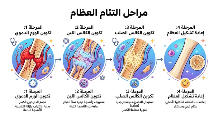
Stages of Bone Healing
1. Inflammation and Hematoma Formation:
- Immediately after the fracture, blood vessels rupture, forming a hematoma (blood clot) at the site.
- This clot provides initial stability and recruits the cells needed for healing, signaling the onset of inflammation.
2. Soft Callus Formation (Fibrocartilaginous):
- Inflammation recruits stem cells that form granulation tissue, then a soft callus (fibrous tissue and cartilage).
- This soft callus begins to bridge the fracture and is often visible on X-ray within a few weeks.
3. Hard Callus Formation (Bony):
- The soft callus is replaced by woven (immature) bone, forming a hard callus.
- This phase involves a change in collagen type (from type II to type I), making the fracture visible and stable.
4. Bone Remodeling:
- This prolonged phase, lasting months to years, refines the bone.
- Woven bone is replaced by stronger lamellar bone, and the medullary canal is reconstituted, restoring the original shape and strength.
5. Clinical Union: (often considered part of remodeling)
- When the callus is fully formed and solid, the bone is considered clinically healed — meaning it can bear weight — even though full remodeling continues.
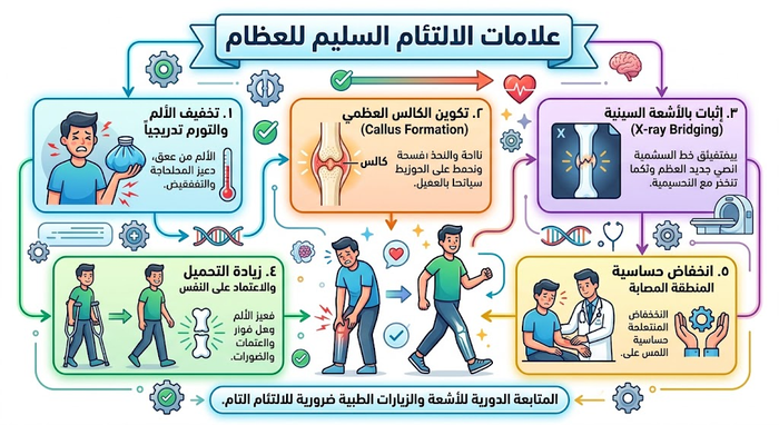
Signs That Your Bone Is Healing Properly
Not sure whether your broken bone is healing as it should? While imaging is the gold standard for tracking bone healing, several clinical signs indicate your body is progressing through the healing stages effectively:
- Gradual decrease in acute pain
- Reduced swelling and bruising
- Ability to move the affected area slightly without sharp pain
- Increasing stability or strength when bearing weight (after your orthopedic surgeon’s clearance)
If you experience severe pain, persistent numbness, or no improvement over time, speak to your doctor. In some cases, delayed healing may require adjustments to the fracture treatment plan.
Bone Fracture Healing Timeline
Bones generally require significant time to heal, typically ranging from several weeks to a few months. On average, most bones heal within 6 to 8 weeks. However, the exact timeline can vary based on the type of fracture and its location.
Factors Affecting Bone Healing
Various factors influence how long the bone healing process takes.
General (Patient) Factors
- Age: Bone healing is typically faster in younger individuals. With advancing age, healing slows due to decreased bone density and slower cell regeneration.
- Nutrition: Proper nutrition is vital. A diet rich in calcium, vitamin D, and protein supports bone formation and repair. Malnutrition can delay healing.
- Smoking: Smoking impairs bone repair by constricting blood vessels. Adequate blood supply is critical for delivering essential nutrients to the fracture site.
- Diabetes: Diabetes can impair circulation and delay bone healing. High blood sugar levels can weaken the function of bone-forming cells.
- Medical conditions: Conditions such as osteoporosis or other chronic diseases can weaken bones, making healing slower and more complicated.
- Medications: Certain medications can interfere with the biological processes of fracture healing and should be monitored by a specialist orthopedic surgeon.
Fracture-Specific Factors
- Fracture type: Simple fractures generally heal faster than complex ones. Comminuted fractures, where the bone shatters into multiple pieces, take longer to heal.
- Location: Healing time varies depending on where the fracture occurs. For example, fractures in areas with good blood supply, such as the ribs, heal faster than those in areas with limited blood flow, such as the tibia.
- Severity: The more severe the fracture, the longer the healing time. Severe fractures may require surgical intervention for proper alignment and fixation.
- Soft tissue injury: Damage to surrounding tissues can impede healing by limiting blood flow and causing additional inflammation.
Last updated: May 2026 — Medically reviewed by Prof. Ibrahim Shaarawi, Lecturer of Orthopedic Surgery, Faculty of Medicine, Ain Shams University.
Frequently Asked Questions
What are the primary biological stages of bone fracture healing?
Secondary (indirect) bone healing proceeds through four overlapping biological phases:
- 1. Hematoma & Inflammatory Phase (Hours to Days): Torn vascular networks bleed into the fracture gap to establish a pro-regenerative hematoma and recruit inflammatory cascades.
- 2. Soft Cartilaginous Callus (Weeks 2 to 3): Fibroblasts and chondroblasts lay down a vascularized fibrocartilaginous bridge across opposing bone ends.
- 3. Hard Bony Callus (Weeks 4 to 12): Endochondral ossification mineralizes the cartilage scaffold into structurally stable, woven bone.
- 4. Bone Remodeling (Months to Years): Continuous osteoclast-osteoblast coupling replaces woven bone with aligned lamellar bone according to functional stress patterns.
What happens during the initial hematoma and inflammatory phase?
Acute skeletal trauma triggers an immediate, localized vascular and immune cascade:
- Hemostasis & Scaffold Construction: Rupture of endosteal, periosteal, and Haversian micro-vessels generates a localized hematoma that coagulates into a provisional fibrin meshwork.
- Cytokine Secretion: De-granulating platelets and infiltrating neutrophils and macrophages release crucial morphogenetic signals, including TGF-β, PDGF, BMPs, and IL-1.
- Cellular Recruitment: These bio-signals create a chemotactic gradient that attracts osteoprogenitor mesenchymal stem cells (MSCs) from adjacent periosteal cambium layers and the bone marrow cavity.
How does a soft callus transform into a hard bony callus?
The transition from a soft to a hard callus occurs via endochondral ossification:
- Chondrocyte Hypertrophy: Chondrocytes inside the soft fibrocartilage enlarge and synthesize type X collagen, simultaneously secreting vascular endothelial growth factor (VEGF).
- Angiogenesis & Matrix Mineralization: Sprouting neo-capillaries penetrate the avascular cartilage, transporting alkaline phosphatase and systemic minerals to initiate calcium phosphate precipitation.
- Osteoblast Deposition: Osteoblasts follow invading vessels, replacing apoptotic chondrocytes with woven bone trabeculae to convert the soft bridge into an immobile, load-resistant hard callus.
What is the difference between primary and secondary bone healing?
The biological pathway of healing is determined by the mechanical strain and stability across the fracture:
- Primary (Direct / Cortical) Healing: Occurs when rigid anatomical internal fixation (compression plating) reduces interfragmentary strain to <2% and eliminates fracture gap motion. Osteoclasts generate cutting cones across the fracture boundary, directly followed by osteoblasts depositing lamellar bone without any visible callus.
- Secondary (Indirect / Callus) Healing: Occurs under relative stability (casts, braces, traction, intramedullary rods) with intermediate strain (2% to 10%). It relies on motion-tolerant endochondral ossification, producing a broad external callus evident on serial radiographs.
How long does the final bone remodeling stage take?
Bone remodeling is the longest phase, typically lasting from 6 months up to several years:
- Structural Refinement: Immature woven bone is biomechanically disorganized; remodeling progressively replaces it with organized, dense lamellar osteons.
- Wolff’s Law: Bone architecture adapts to applied mechanical loading. Bone is added along dominant stress vectors and resorbed where mechanical forces are minimal.
- Canal Recanalization: Osteoclasts hollow out the hard callus envelope and excavate the central intramedullary cavity, restoring normal diaphyseal marrow architecture and vascular patency.
What systemic and local factors delay or prevent bone healing (nonunion)?
Failure of bone consolidation can arise from localized mechanical issues or systemic metabolic deficits:
- Mechanical Instability: Insufficient immobilization causes excessive interfragmentary motion, shearing newly formed capillary networks and driving pseudoarthrosis (false joint formation).
- Vascular Deprivation: Extensive soft-tissue stripping or high-energy trauma deprives bone margins of blood flow, producing avascular bone ends.
- Nicotine & Tobacco Exposure: Causes peripheral vasoconstriction, reduces endothelial cell proliferation, and decreases oxygen delivery by up to 50%.
- Metabolic Factors: Uncontrolled diabetes mellitus (advanced glycation end-products impair osteoblast activity), malnutrition, hyperparathyroidism, and severe hypovitaminosis D.
Why do pediatric bone fractures heal faster than adult fractures?
Skeletal physiology in pediatric patients offers distinct regenerative advantages:
- Hypervascular Periosteum: Children possess a thick, robust, osteogenic periosteum that frequently remains intact on one side of a fracture (hinge effect), providing an immediate vascular conduit and osteoblast reservoir.
- Accelerated Callus Consolidation: High metabolic activity and abundant stem cell populations routinely allow pediatric long-bone fractures to achieve clinical union in 3 to 6 weeks, compared to 8 to 12 weeks in mature adults.
- High Remodeling Capacity: Open growth plates (physes) continuously generate skeletal length, allowing spontaneous realignment of angular deformities, provided the fracture is close to a physis and aligned in the joint’s plane of motion.
What are the clinical and radiographic criteria for a fully healed fracture?
Orthopedic surgeons confirm complete bony union through a combined diagnostic evaluation:
- Clinical Union: Complete resolution of pain and tenderness upon direct palpation of the original fracture line, absence of gross or micro-motion during manual stress testing, and painless full weight-bearing ambulation.
- Radiographic Union: Progressive disappearance of the radiolucent fracture line and the presence of continuous bridging bone trabeculae across at least 3 out of 4 cortices on orthogonal (anteroposterior and lateral) plain X-ray views.
What nutritional interventions accelerate fracture repair?
Bone repair places substantial metabolic and cellular demands on nutritional reserves:
- Dietary Protein: Requires 1.2 to 1.5 g/kg/day. Protein supplies essential amino acids (proline, lysine, glycine) necessary for building the extracellular type I collagen framework.
- Calcium & Vitamin D: Daily intake of 1,000 to 1,200 mg elemental calcium paired with adequate vitamin D3 (targeting serum 25(OH)D levels >30 ng/mL) ensures substrate availability for hard callus mineralization.
- Ascorbic Acid (Vitamin C): Acts as an essential cofactor for prolyl and lysyl hydroxylases; deficiency destabilizes triple-helix collagen cross-linking.
- Micronutrients: Zinc stimulates alkaline phosphatase activity, and magnesium supports calcium metabolism and osteoblast proliferation.
Can nonsteroidal anti-inflammatory drugs (NSAIDs) interfere with fracture healing?
Yes, high-dose NSAIDs should be used cautiously during early fracture management:
The early inflammatory reaction is an obligatory trigger for bone repair. Infiltrating inflammatory cells rely on cyclooxygenase-2 (COX-2) enzymes to produce prostaglandin E2 (PGE2), which recruits osteoprogenitor cells and initiates vascular ingrowth.
Nonsteroidal anti-inflammatory drugs (e.g., ibuprofen, diclofenac, naproxen) and selective COX-2 inhibitors inhibit this pathway. Animal models and clinical studies link high-dose, continuous NSAID use during the first 2 to 4 weeks post-injury with delayed callus formation and elevated nonunion rates. Orthopedic guidelines recommend using acetaminophen (paracetamol) or short courses of mild opioids as first-line alternatives for acute pain management.
References
AAOS OrthoInfo — Fractures (Broken Bones) (2024)
https://orthoinfo.aaos.org/en/diseases–conditions/fractures-broken-bones/- AAOS OrthoInfo — Helping Fractures Heal (Orthobiologics) (2024)
https://orthoinfo.aaos.org/en/treatment/helping-fractures-heal-orthobiologics/ - Cleveland Clinic — Bone Fractures: Types, Symptoms & Treatment (2024)
https://my.clevelandclinic.org/health/diseases/15241-bone-fractures - Cleveland Clinic — Malunion and Nonunion Fractures (2024)
https://my.clevelandclinic.org/health/diseases/malunion-nonunion-fracture - Mayo Clinic — Fractures (Broken Bones): First Aid (2024)
https://www.mayoclinic.org/first-aid/first-aid-fractures/basics/art-20056641 - NICE — Fractures (Non-Complex): Assessment and Management (NG38) (2023)
https://www.nice.org.uk/guidance/ng38 - ScienceDirect — Fracture Nonunion in Long Bones: A Literature Review of Risk Factors and Surgical Management (2020)
https://www.sciencedirect.com/science/article/pii/S0020138320309554 - PubMed — Delayed Tibial Shaft Fracture Healing Associated with Smoking: Systematic Review and Meta-Analysis (2021)
https://pubmed.ncbi.nlm.nih.gov/34639529/