
Quick answer
Enchondroma is a benign cartilage tumour that grows inside a bone, most often in the finger bones and then the femur and humerus. Most are found by chance on an X-ray or MRI done for something else, and they need monitoring rather than surgery. We operate (curettage and bone grafting) when one causes pain, a fracture or worrying changes.
✅ Last medically reviewed by Prof. Dr. Ibrahim Shaarawi, Professor of Orthopaedic Surgery, on 27 October 2026
People often arrive worried by one line in a knee or shoulder MRI report: “cartilaginous lesion in the medullary cavity”. Most of the time this is an enchondroma that only needs follow-up. This guide explains when we can relax and when we act. It is part of our bone and soft tissue tumor surgery guide.
What is an enchondroma?
It starts from a small island of cartilage left inside the bone and grows slowly. It can appear at any age and is most common in middle age (OrthoInfo). Typical sites:
- The finger bones and metacarpals of the hand.
- The lower end of the femur, at the knee.
- The upper humerus, at the shoulder.
Reports may use the term atypical cartilaginous tumour, a closely related category for long-bone cartilage tumours that cannot be told apart on imaging alone. An enchondroma should not be confused with an osteochondroma, which grows outward from the bone surface.
How it is found
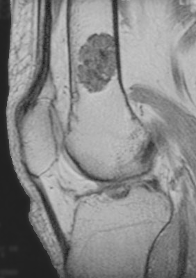
- By chance: the most common route, on a knee or shoulder MRI for another problem such as a meniscus tear.
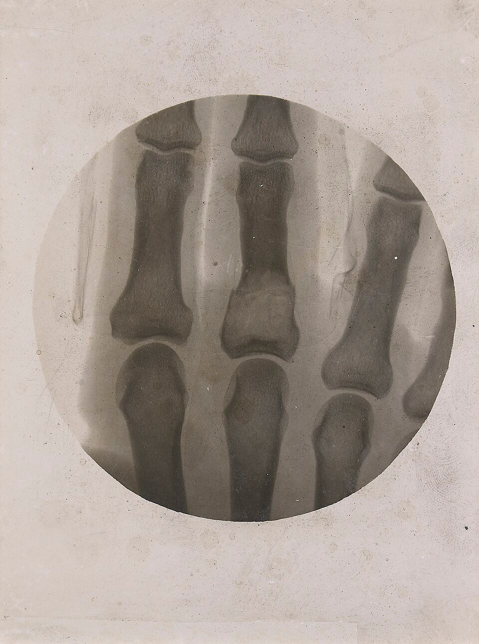
- Finger swelling: the tumour slowly expands the small bone.
- A fracture after minor injury: the tumour thins the cortex of the finger bone.
If knee or shoulder pain has another clear cause, the enchondroma is usually not the source. That matters before anyone considers surgery.
Is it dangerous? Enchondroma versus chondrosarcoma
A single enchondroma rarely becomes malignant (OrthoInfo). The difficulty is that a low-grade chondrosarcoma can look similar. Signs that make us reassess or biopsy:
- Pain at the tumour itself, especially at night, without a fracture.
- Size over 5 cm in a long bone.
- Deep scalloping of the inner cortex or a break in the cortex.
- Growth between two scans.
- Multiple tumours (Ollier disease or Maffucci syndrome).
MRI is the key test; see MRI for bone tumors. The lesion may also light up on a bone scan for bone tumors, which on its own does not mean it is malignant.
Video: Treating a finger tumour and other benign tumours
Key numbers to know
- Cartilage tumours were found by chance on knee MRI in 2.8% of 1,285 people, with a mean size of 12 mm (Stomp et al., 2015). In a review of 44,762 knee MRIs the rate was 1.45%, and 72.9% sat in the lower femur (Woltsche et al., 2023).
- On MRI follow-up of 128 cartilage tumours in long bones over a mean of 50 months, 87% stayed the same or shrank, and none became a high-grade chondrosarcoma (Deckers et al., 2021).
- Of 102 hand enchondromas treated with curettage, 93% did not recur and 61% healed completely on X-ray at a median of 6 months (Sassoon et al., 2012).
- In Ollier disease and Maffucci syndrome (multiple enchondromas), 40% of patients developed a chondrosarcoma, with a higher risk when the pelvis was involved (Verdegaal et al., 2011).
| Treatment | When we choose it | What to expect |
|---|---|---|
| X-ray or MRI surveillance | A painless lesion in a long bone that looks quiet on MRI | Most lesions stay unchanged for years |
| Curettage and bone grafting | A hand lesion causing pain, thinning of the bone or fracture | The bone heals over months with a low recurrence rate |
| Fracture care first, then curettage | A finger fracture through the tumour | The fracture heals, then the tumour is removed in a planned operation |
| Biopsy, then wider surgery | Night pain, growth or erosion of the bone cortex | Rules out a low-grade chondrosarcoma |
At Prof. Dr. Ibrahim Shaarawi’s clinics in Nasr City, Mohandeseen and New Cairo (5th Settlement) we look at the images themselves, not just the report, explain what they show, and agree with you on follow-up or the right operation.
Treatment: monitor or operate?
Most surgeons do not remove enchondromas that cause no symptoms (OrthoInfo). We follow them with X-ray or MRI, set the intervals by site and size, and space them out when the lesion stays the same. Surgery means curettage, scraping the tumour out, then filling the cavity with bone graft or a bone substitute.
When a finger fractures through the tumour, we can let the fracture heal first and then curette, or treat both together; outcomes did not differ between the two in a series of 102 hand enchondromas (Sassoon et al., 2012).
What to do if it appears on your report
- Do not rush into surgery or a biopsy before a bone tumour surgeon has seen the images themselves.
- Keep the CD of every MRI; comparing two scans is what reassures us.
- Tell your doctor about any new night pain at the site.
- Compare with related benign lesions such as giant cell tumor of bone, which behaves very differently.
Frequently asked questions about enchondroma
Is an enchondroma cancer?
No, it is benign. Malignant change into chondrosarcoma is rare for a single lesion and more likely when there are many.
Does it have to be removed?
Not always. Most painless lesions are only monitored; we remove them for pain, fracture or worrying MRI features.
Will it go away on its own?
It usually does not disappear, but most stay unchanged for years and some shrink slightly on MRI.
Do I need a biopsy?
Not for a lesion that looks quiet on imaging. A biopsy is reserved for worrying signs and is planned by the tumour surgeon.
How long does the bone take to heal after curettage?
In the hand the bone usually heals over a few months, and finger movement returns gradually with exercises.
To have your scans reviewed by a professor of orthopaedic oncology, book a consultation, call +20 155 240 4488 or message us on WhatsApp at +20 155 554 7181. Bring the images on CD, not only the report.
Medically reviewed by Prof. Dr. Ibrahim Shaarawi, Professor of Orthopaedic Surgery. This article is general medical education and does not replace an examination or the plan your own doctor sets for you. See our medical disclaimer.
Related bone and soft tissue tumor guides
Related guides on benign bone tumours and their tests:
- Bone and soft tissue tumor surgery
- MRI for bone tumors
- Bone scan for bone tumors
- Giant cell tumor of bone
References
- American Academy of Orthopaedic Surgeons (OrthoInfo). Enchondroma.
- Ahlawat S, et al. ACR Appropriateness Criteria® Suspected Primary Bone Tumors: 2024 Update. J Am Coll Radiol. 2025;22(5S):S440-S454.
- Stomp W, et al. Prevalence of cartilaginous tumours as an incidental finding on MRI of the knee. Eur Radiol. 2015;25(12):3480-7.
- Deckers C, et al. Midterm MRI Follow-Up of Untreated Enchondroma and Atypical Cartilaginous Tumors in the Long Bones. Cancers (Basel). 2021;13(16).
- Sassoon AA, et al. Enchondromas of the hand: factors affecting recurrence, healing, motion, and malignant transformation. J Hand Surg Am. 2012;37(6):1229-34.
- Verdegaal SH, et al. Incidence, predictive factors, and prognosis of chondrosarcoma in patients with Ollier disease and Maffucci syndrome: an international multicenter study of 161 patients. Oncologist. 2011;16(12):1771-9.
- Woltsche JN, et al. Prevalence and characteristics of benign cartilaginous tumours of the knee joint as identified on MRI scans. Cancer Imaging. 2023;23(1):50.