
Giant cell tumor of bone (GCTB) is a benign but locally aggressive tumour that attacks the ends of long bones, most often around the knee and wrist. It is entirely different from giant cell tumour of the tendon sheath, which involves soft tissues of the hand. Modern treatment relies on extended curettage, cavity filling, and — in selected cases — the biological drug denosumab, with the goal of clearing the tumour while preserving the joint.
What is giant cell tumor of bone?
GCTB is built from multinucleated osteoclast-like giant cells that erode healthy bone. It is classified as benign, yet it behaves aggressively at its site: left untreated it destroys the affected bone and can breach the neighbouring joint. It appears mostly in adults between 20 and 40 years old, is slightly more common in women, and clusters at the ends of long bones — the distal femur and proximal tibia around the knee, and the distal radius at the wrist.
Warning signs
- Persistent pain that worsens with movement or weight-bearing and does not respond to ordinary analgesics.
- A firm swelling or palpable mass around the affected joint.
- Pathological fracture — the bone breaks after trivial force because the tumour has hollowed it out.
- Restricted joint motion — difficulty bending or straightening the nearby joint.
GCTB versus tenosynovial giant cell tumor
Patients often confuse GCTB with the similarly named tumour of the tendon sheath. The distinction is anatomical rather than semantic. GCTB grows inside bone at the knee or wrist, behaves aggressively, and needs extended curettage with cement or bone graft. Tenosynovial GCT grows in the soft tissue around finger tendons, presents as a slow-growing painless nodule, and is treated with precise marginal excision to protect the underlying tendon.
Diagnosis
Assessment starts with plain X-rays, which typically show a lytic (radiolucent) lesion at the end of a long bone extending to the joint surface. MRI defines the tumour’s real extent, its relationship to the joint cartilage, and the surrounding neurovascular structures. A chest CT is done before surgery to look for the small proportion of GCTB cases that seed benign nodules to the lung. The definitive diagnosis is histological, and the biopsy tract must be planned by the surgeon who will do the definitive resection — a poorly placed biopsy tract can convert a joint-preserving operation into a wide resection.
Modern treatment protocol
Amputation is no longer part of the standard algorithm. Three complementary strategies dominate current practice:
- Extended intralesional curettage — the entire tumour is scraped from inside the bone, the cavity walls are polished with a high-speed burr, and a local adjuvant such as phenol, liquid nitrogen, or electrocautery is applied to kill microscopic residual cells.
- Cavity filling — polymethyl methacrylate (PMMA) cement is the most common choice because it delivers immediate mechanical support, and its curing heat helps sterilise the last millimetres of bone. Autograft or processed bone-bank graft is preferred when biological reconstruction is important, particularly in younger patients.
- Denosumab — a targeted RANKL inhibitor used for tumours too large or too close to critical structures for primary curettage. It hardens the tumour and shrinks it enough to permit later surgery, but is used with caution because preoperative use has been associated with a higher local recurrence rate after subsequent curettage.
How the surgeon chooses the plan
The decision follows a stepwise logic. Small tumours away from cartilage receive extended curettage with cement. Larger tumours abutting the joint surface add reinforcing plates and screws. Tumours that have breached the joint or recurred repeatedly are treated by wide resection and reconstruction with a tumour endoprosthesis. Denosumab is reserved for the tumours where surgery would otherwise sacrifice a joint, and is not offered routinely for straightforward cases.

Rehabilitation timeline
Recovery moves through four phases: protection with crutches for the first fortnight, restoration of joint range from weeks two to six, strengthening and progressive loading from six weeks to three months, and return to daily life beyond the third month. High-impact activity — running, jumping, contact sport — remains restricted until the surgeon confirms that bone density has been re-established. Physiotherapy is not optional; because the cement does not integrate biologically with bone, the muscles around the joint have to compensate, and their strength defines the long-term function of the limb.
Recurrence and follow-up
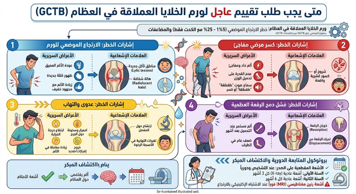
GCTB recurs in a meaningful minority of cases even after correctly performed extended curettage, and recurrence almost always shows up within the first two years. Follow-up X-rays every three months during year one and every six months during year two are non-negotiable. New pain, a growing swelling, or a widening lucent halo between bone and cement is an indication for immediate MRI. Because GCTB can seed benign lung nodules, a chest CT is added to the surveillance schedule.
When to seek urgent assessment
Book an urgent orthopaedic oncology appointment for persistent night pain lasting more than four weeks, a growing lump near the knee or wrist, or a fracture after minor injury. Sudden pain with inability to bear weight, or new swelling after a previous surgery, should be treated as an emergency.
Frequently Asked Questions
What is a Giant Cell Tumor of Bone (GCTB) and is it considered malignant?
A Giant Cell Tumor of Bone (GCTB) is an osteolytic neoplasm arising predominantly in the epiphyseal region of mature long bones. Histologically, it is classified as benign but locally aggressive, meaning it exhibits significant capacity for bone resorption and local soft tissue infiltration:
- Benign Nature: More than 95% of cases show typical non-malignant histology and do not undergo systemic transformation.
- Malignant Transformation: Primary or secondary malignant GCTB (dedifferentiation into high-grade osteosarcoma or undifferentiated pleomorphic sarcoma) occurs in less than 2% of patients.
- Benign Pulmonary Implants: Approximately 1% to 3% of patients develop histologically benign lung nodules that follow an indolent clinical course.
Which age group is most susceptible and where do these tumors typically develop?
GCTB displays distinct epidemiological and anatomical characteristics:
- Age Group: It peaks between 20 and 45 years of age, occurring almost exclusively after growth plate (physeal) closure. It is rare in skeletally immature pediatric patients.
- Gender Predilection: There is a slight female predominance (approximately 1.2:1).
- Predominant Anatomical Locations:
- Around the knee (50%–60% of cases): The distal femur and proximal tibia represent the most common sites.
- Distal radius: The second most frequent long bone location (approx. 10%), often displaying higher local recurrence rates.
- Proximal humerus: Shoulder region.
- Axial skeleton: The sacrum and vertebral bodies, representing surgically challenging locations.
What are the classic symptoms and signs of Giant Cell Tumor of Bone?
Symptoms develop insidiously as the tumor hollows out structural trabeculae beneath the joint surface:
- Persistent, Deep Bone Pain: Discomfort localized adjacent to the affected joint, which worsens with mechanical weight-bearing and physical exertion, progressing over time to rest and nocturnal pain.
- Palpable Swelling or Mass: A firm, tender expansion over the bone contour with localized skin warmth due to increased tumor vascularity.
- Joint Stiffness & Effusion: Limited terminal flexion or extension with secondary reactive joint fluid accumulation.
- Pathological Fracture: Presenting symptom in 10% to 30% of patients, occurring during routine activities as the structural cortical shell collapses.
What is the Campanacci grading system used to stage GCTB?
The Campanacci staging system is the international radiographic and surgical benchmark used to stratify local disease extent and determine operative strategy:
- Grade I (Latent / Inactive): Small, intraosseous lesion with well-demarcated margins, a thin rim of sclerotic bone, and an intact cortical shell.
- Grade II (Active): Moderately large, expansile osteolytic lesion causing marked cortical thinning without a sclerotic rim, but the periosteal envelope remains intact (most common presentation).
- Grade III (Aggressive): Rapidly expanding lesion showing complete cortical destruction, breakthrough of the periosteal barrier, and extension into adjacent soft tissue compartments or intra-articular structures.
How is Giant Cell Tumor diagnosed and why is a biopsy mandatory?
Accurate diagnosis requires a multimodal staging and histological pathway:
- Plain Radiographs (X-ray): Demonstrates an eccentric, purely geographic lytic lesion originating in the epiphyseal/metaphyseal region and extending down to the subchondral plate, with a classic ‘soap-bubble’ trabecular appearance without tumor matrix mineralization.
- Magnetic Resonance Imaging (MRI): Essential to define the exact extent of intraosseous spread, joint cartilage integrity, and soft tissue margins, typically showing low-to-intermediate T1 and heterogeneous T2 signal.
- Computed Tomography (CT): Evaluates residual cortical thickness, fractures, and subchondral bone support.
- Core Needle Biopsy: Mandatory prior to any definitive intervention to demonstrate uniformly distributed, multinucleated osteoclast-like giant cells embedded within mononucleated stromal cells, and to definitively rule out osteosarcoma, brown tumors of hyperparathyroidism, or aneurysmal bone cysts (ABCs).
What is extended intralesional curettage and how does it spare the joint?
Extended intralesional curettage is the oncological gold standard for joint preservation in Campanacci Grade I and II lesions:
- Cortical Window: A generous cortical fenestration is cut over the tumor to provide complete visual access to all margins of the bone cavity.
- Gross Debridement: The vascular, fleshy tumor tissue is scooped out with specialized angled curettes.
- High-Speed Burr Drilling: A high-speed surgical burr is systematically applied to grind away an additional 2 to 3 mm of the peripheral cancellous walls, eliminating microscopic cellular extensions hidden within the bone trabeculae.
- Reconstruction & Fixation: The void is filled with polymethylmethacrylate (PMMA) bone cement, which provides immediate structural rigidity, often reinforced with locked plates and screws to prevent postoperative fracture and enable immediate weight-bearing.
Which local chemical and thermal adjuvants are used during surgery?
Simple curettage alone yields unacceptable local recurrence rates approaching 50%. The routine addition of intraoperative local adjuvants drops recurrence to 10%–15%:
- Medical Phenol (70%): A chemical agent painted onto the cavity walls to induce protein coagulation and cell necrosis in microscopic tumor residuals, followed by an absolute alcohol wash for neutralization.
- Liquid Nitrogen Cryotherapy: Controlled sub-zero freezing cycles produce intracellular ice crystal formation and rapid cell lysis within the peripheral margin.
- Hydrogen Peroxide (3%): Provides mechanical cleansing, local hemostasis, and oxidative cytotoxicity.
- PMMA Thermal Cautery: The exothermic polymerization reaction of PMMA bone cement reaches temperatures between 60°C and 80°C, producing a localized thermal burn that kills residual tumor cells in the adjacent bone bed.
When is wide en-bloc resection with megaprosthetic reconstruction necessary?
Joint-sparing curettage is not feasible in all clinical scenarios. Wide en-bloc resection is indicated when:
- The lesion is an aggressive Campanacci Grade III tumor with complete collapse and destruction of the subchondral bone and articular surface.
- There is a comminuted, displaced intra-articular pathological fracture where anatomical joint reconstruction is impossible.
- The patient experiences multiple local recurrences with extensive joint and soft tissue compromise.
- The tumor involves an anatomically expendable bone segment (such as the proximal fibula or distal ulna) that can be removed with zero functional deficit.
- Reconstruction involves implanting a modular tumor megaprosthesis (e.g., distal femoral or proximal tibial replacement) or structural osteoarticular allografts to restore limb stability and mobility.
What is the role of Denosumab in treating Giant Cell Tumor of Bone?
Denosumab (Xgeva) is a fully human monoclonal antibody that specifically targets and inhibits RANKL, disrupting the signaling pathway that drives the formation and survival of osteoclast-like giant cells:
- Neoadjuvant Therapy: Administered for 3 to 6 months prior to surgery to halt bone osteolysis, induce a firm osteosclerotic rim, and shrink soft tissue components, converting difficult lesions into candidates for joint-preserving curettage.
- Surgically Inaccessible Lesions: Primary management for tumors located in the sacrum, spine, or skull base where complete surgical resection risks unacceptable neurological morbidity.
- Metastatic & Recurrent Disease: Long-term disease control for recurrent unresectable lesions and benign lung metastases.
- Clinical Precaution: Patients require routine monitoring of serum calcium to prevent hypocalcemia, alongside dental evaluations to mitigate the risk of medication-related osteonecrosis of the jaw (MRONJ).
What is the local recurrence rate and what follow-up protocol is required?
While recurrence rates after extended curettage with adjuvants and cementation remain low (10% to 15%), over 80% of local recurrences occur within the first 24 to 36 months. Strict oncologic surveillance is required:
- Years 1 to 2: Clinical evaluations and orthogonal plain radiographs of the operated bone every 3 to 4 months to assess cement-bone interfaces for radiolucent lines indicating recurrence.
- Years 3 to 5: Clinical and radiographic check-ups every 6 months.
- Pulmonary Monitoring: Plain chest X-rays or low-dose chest CT scans every 6 to 12 months for 5 years to screen for asymptomatic benign lung metastases.
References
المراجع العلمية الدولية
- American Academy of Orthopaedic Surgeons – OrthoInfo. Giant Cell Tumor of Bone. orthoinfo.aaos.org
- Johns Hopkins Medicine. Giant Cell Tumor. hopkinsmedicine.org
- Errani C, Tsukamoto S, Leone G, et al. Denosumab may increase the risk of local recurrence in patients with giant-cell tumor of bone treated with curettage. Journal of Bone and Joint Surgery. journals.lww.com
- Zhao Y, Cai Z, Tang X, et al. Preoperative Denosumab may increase the Risk of Local Recurrence of Giant-cell Tumor of Bone Treated with Curettage: A Systematic Review and Meta-analysis. Journal of Cancer. jcancer.org
- Bone Cancer Research Trust. Giant Cell Tumour of the Bone — Information for Healthcare Professionals. bcrt.org.uk
- Radiopaedia. Giant cell tumour of bone. radiopaedia.org
- NICE Clinical Knowledge Summaries. Bone and soft tissue sarcoma: recognition and referral . cks.nice.org.uk