
Quick answer
Osteochondroma is a benign bony outgrowth with a cartilage cap that grows from near a growth plate, usually around the knee or shoulder, in children and teenagers. It is the most common benign bone tumour. Most need only observation and stop growing when the child stops growing. We remove it when it causes pain, rubs a tendon, presses on a nerve or vessel, or grows after puberty. Some children have many (multiple hereditary exostoses), which needs closer follow-up.
✅ Last medically reviewed by Prof. Dr. Ibrahim Shaarawi, Professor of Orthopaedic Surgery, on 28 October 2026
Parents usually find the lump by chance, often while bathing a child or after a knock. The first worry is cancer. In almost every case it is not, and the real questions are whether it needs removing and when. This article is part of our guide to bone spurs.
What is an osteochondroma?
Long bones grow from cartilage plates near their ends. In an osteochondroma a small piece of that plate grows sideways, forming bone covered by a cartilage cap. It grows while the child grows and normally stops at the end of puberty. The American Academy of Orthopaedic Surgeons lists it as the most common benign bone tumour (OrthoInfo).
- Solitary osteochondroma: a single lump, the usual form.
- Multiple hereditary exostoses: many lumps, usually inherited through the EXT1 or EXT2 genes, and often diagnosed in early childhood.
Symptoms
- A hard, painless lump near the knee, shoulder or ankle.
- Pain with activity when a tendon or muscle rubs over it, sometimes with a snapping feeling.
- Numbness or tingling if it presses on a nerve, such as behind the knee.
- Rarely, a change in pulse or colour if it presses on a blood vessel.
- Sudden pain if the stalk breaks after a fall.
Children with multiple exostoses can also develop a short forearm, a bowed leg or a difference in leg length, because the lumps disturb normal growth.
Video: Osteochondroma in children
Diagnosis
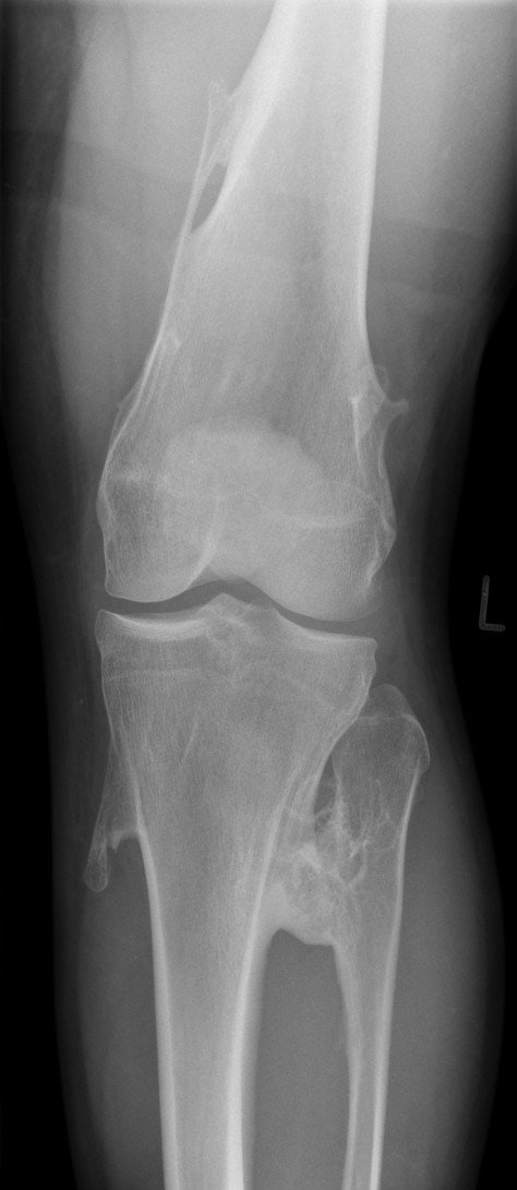
A plain X-ray usually shows the typical shape: a bony stalk or bump continuous with the bone it grows from. We order an MRI when we need to measure the cartilage cap or see the nerves and vessels close to it. A cap that is thick in an adult can be a warning sign, so we compare it over time.
Multiple hereditary exostoses
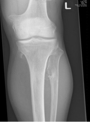
About 70% of cases are inherited from a parent and 30% appear for the first time in the child (OrthoInfo). These children often need several operations during growth, either to remove painful lumps or to correct a bowed forearm or leg. We follow them until growth ends and teach the family which changes need an earlier visit. Related growth problems are covered in enchondroma.
Key numbers to know
- A solitary osteochondroma is the most common benign bone tumour, making up 35 to 40% of benign bone tumours. The lifetime risk of it turning malignant is under 1% for a single lesion and about 10% in multiple hereditary exostoses (AAOS OrthoInfo).
- Multiple osteochondromas affect about 1 in 50,000 people and are slightly more common in males (Bovée, 2008).
- In a survey of 779 people with multiple hereditary exostoses, patients had a mean of 7.3 operations each, and 2.7% developed malignant change at a mean age of 28, most often in the pelvis (Czajka and DiCaprio, 2015).
| Treatment | When we choose it | What to expect |
|---|---|---|
| Observation | A small painless lesion not pressing on a nerve or vessel | An X-ray if pain or growth appears, and review until growth ends |
| Excision | Pain with activity, tendon snapping, pressure on a nerve or artery, or an unwanted bump | A small incision and usually back to school within days; recurrence is rare if the cartilage cap is removed |
| Excision plus deformity correction | Multiple lesions causing a short forearm or a bowed leg | Realigning the bone or guiding growth depending on age |
| Biopsy, then wider surgery | New pain, growth after puberty, or a thick cartilage cap on MRI | Ruling out change into a chondrosarcoma |
At Prof. Dr. Ibrahim Shaarawi’s clinics in Nasr City, Mohandeseen and New Cairo (5th Settlement) we examine your child, compare the new X-ray with earlier ones, and agree with you on watching or the right time to remove it.
Treatment
Most solitary osteochondromas need no operation. We see the child, take an X-ray when there is pain or a change in size, and review until growth is complete. We recommend removal when:
- it hurts with activity or interferes with sport or school;
- it rubs on a tendon or presses on a nerve or vessel;
- it causes a deformity or limits joint movement;
- it grows or becomes painful after growth has finished, which needs assessment first.
In surgery we remove the lump at its base together with its cartilage cap and periosteum. Leaving part of the cap is the main reason for regrowth. Most children go home the same or the next day and return to school within days, with sport after a few weeks depending on the site.
Can it turn into cancer?
Rarely. The risk is under 1% for a solitary lesion and higher in multiple exostoses (OrthoInfo). Warning signs in an adult are new pain, a lump that grows, or a thick cartilage cap on MRI. These are reasons for imaging and, if needed, a planned biopsy by a tumour surgeon. Our approach to bone tumours is described in bone and soft tissue tumor surgery.
Frequently asked questions about osteochondroma
Is osteochondroma cancer?
No. It is a benign growth. The lifetime risk of malignant change is under 1% for a single lesion and about 10% in multiple hereditary exostoses.
Does every osteochondroma need surgery?
No. Most are watched. We remove them when they cause pain, rub a tendon, press on a nerve or vessel, or cause deformity.
Will it grow back after removal?
Regrowth is uncommon when the cartilage cap is removed completely; it is more likely if surgery is done very early in a young child.
Can my child play sport?
Usually yes. If the lump is in a spot that gets knocked or rubs a tendon, removal may be advised so the child can play without pain.
Is multiple hereditary exostoses inherited?
About 70% of cases are inherited from a parent, and each child of an affected parent has a high chance of having the condition.
To have your foot, heel or knee checked by Prof. Dr. Ibrahim Shaarawi, book a consultation, call +20 155 240 4488 or message us on WhatsApp at +20 155 554 7181. Bring any previous X-rays with you.
Medically reviewed by Prof. Dr. Ibrahim Shaarawi, Professor of Orthopaedic Surgery. This article is general medical education and does not replace an examination or the plan your own doctor sets for you. See our medical disclaimer.
Related bone spur guides
Related guides:
References
- American Academy of Orthopaedic Surgeons (OrthoInfo). Osteochondroma.
- Bovée JV. Multiple osteochondromas. Orphanet J Rare Dis. 2008;3:3.
- Czajka CM, DiCaprio MR. What is the Proportion of Patients With Multiple Hereditary Exostoses Who Undergo Malignant Degeneration?. Clin Orthop Relat Res. 2015;473(7):2355-61.
- Alabdullrahman LW, et al. Osteochondroma. In: StatPearls. Treasure Island (FL): StatPearls Publishing; 2024.