
Quick answer
Synovial chondromatosis is a benign condition in which part of the joint lining turns into cartilage, forming small nodules that break off and float in the joint as loose bodies. It mostly affects the knee, then the hip, shoulder and elbow, causing pain, swelling and locking. Treatment removes the loose bodies together with the diseased lining, and arthroscopy is enough in most cases.
✅ Last medically reviewed by Prof. Dr. Ibrahim Shaarawi, Professor of Orthopaedic Surgery, on 27 October 2026
Some patients say their knee feels “full of gravel”, and that is close to what we find: dozens of cartilage bodies, some the size of a lentil or bigger. This guide covers diagnosis and explains why removing the loose bodies alone is not enough. It is part of our bone and soft tissue tumor surgery guide.
What is synovial chondromatosis?
Nodules of cartilage form inside the synovium, grow, then detach into the joint fluid. Over time many calcify and become visible on X-ray. AAOS separates two forms (OrthoInfo):
- Primary: a rare benign tumour arising from the lining itself, and the subject of this article.
- Secondary: loose bodies caused by arthritis or an old injury. Not a tumour; treatment depends on the cause.
Symptoms
- Joint pain that worsens with activity.
- Recurrent swelling and fluid.
- Stiffness and gradual loss of movement.
- Sudden locking, clicking or grinding.
- Sometimes small mobile lumps can be felt around the knee.
It usually appears between 30 and 50, is more common in men, and rarely involves more than one joint. Because it feels like arthritis, diagnosis can be delayed for years.
Diagnosis on X-ray and MRI
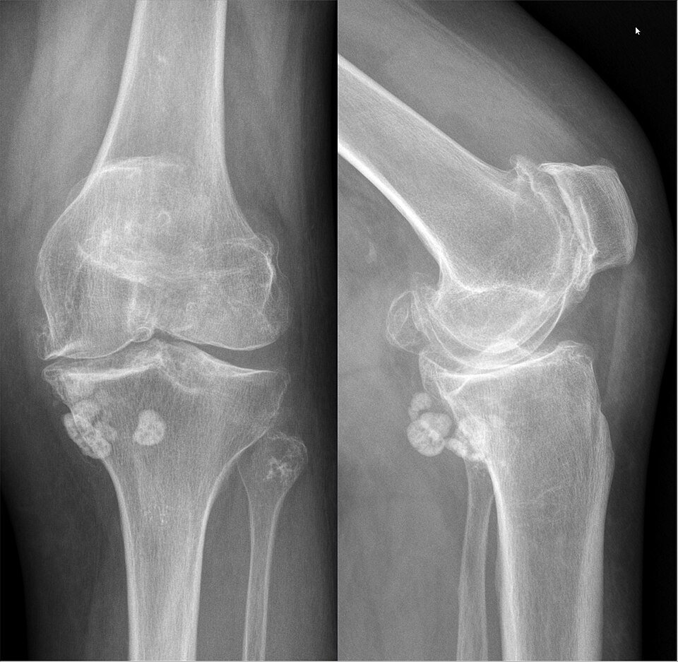
Calcified bodies are easy to see on X-ray and tend to be similar in size. Non-calcified bodies are invisible on X-ray, so MRI or CT is needed (Murphey et al., 2007). MRI also shows how much lining is affected and the state of the cartilage, both of which change the surgical plan.
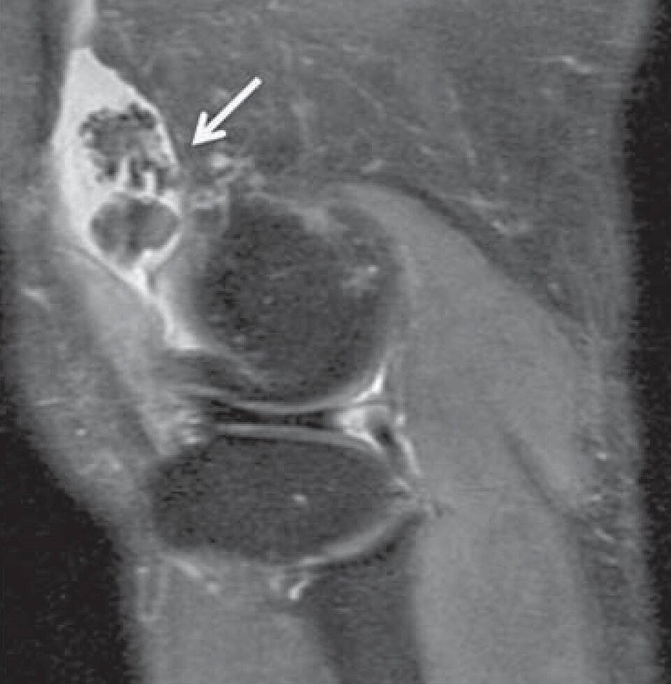
We distinguish it from pigmented villonodular synovitis (PVNS), which does not form calcified bodies and looks dark on MRI, and from loose bodies caused by arthritis. See also MRI for bone tumors. The video below is from an arthroscopic synovectomy of the knee, the same technique we use to clear cartilage bodies and diseased lining.
Video: Arthroscopic removal of a synovial tumour (PVNS) of the knee
Key numbers to know
- In a systematic review of arthroscopic treatment for knee synovial chondromatosis, the condition returned in 22.6% of patients, mostly after loose-body removal without synovectomy (Alamiri et al., 2025).
- In a review of 120 patients, recurrence was 5.7% after arthroscopic synovectomy, 31.4% after arthroscopic washout alone and 12.5% after open synovectomy (Liang et al., 2025).
- In the hip, 197 patients across 14 studies had arthroscopic surgery, with a 7.1% recurrence rate and 1% minor complications (de Sa et al., 2014).
| Treatment | When we choose it | What to expect |
|---|---|---|
| Observation | Mild symptoms and a healthy joint | Periodic review and X-rays to watch for arthritis |
| Arthroscopic loose body removal | A few loose bodies and inactive synovium | Quick relief, but higher recurrence if diseased synovium is left |
| Loose body removal plus arthroscopic synovectomy | Most knee and hip cases | The lowest recurrence rates in recent reviews |
| Open surgery | Very large bodies or areas the arthroscope cannot reach | Complete clearance with longer recovery |
| Joint replacement | Advanced arthritis in an older patient | Treats pain and arthritis together |
At Prof. Dr. Ibrahim Shaarawi’s clinics in Nasr City, Mohandeseen and New Cairo (5th Settlement) we look at the images themselves, not just the report, explain what they show, and agree with you on follow-up or the right operation.
Treatment: why loose body removal alone falls short
The loose bodies cause the locking and pain, but the lining produces them. Remove the bodies and leave the diseased synovium, and new ones form. That is why most recurrences in recent reviews followed loose body removal alone, and the lowest recurrence followed removal plus arthroscopic synovectomy (Liang et al., 2025).
- Arthroscopy: first choice for most knees and hips, clearing every body and the affected lining.
- Open surgery: for very large bodies or areas the arthroscope cannot reach.
- Joint replacement: for older patients with advanced arthritis, treating pain and arthritis together.
For more on the technique, see knee arthroscopy.
Can it turn malignant?
Change into chondrosarcoma is very rare: a systematic review found only 48 published cases since 1957 (Ng et al., 2017). Signs that make us biopsy and reassess:
- Rapid return after complete surgery.
- Pain and swelling that keep getting worse over months.
- Spread into the bone marrow on MRI.
A biopsy in that setting should follow a track planned by the tumour surgeon, not a random needle.
After surgery and follow-up
After arthroscopy most patients walk the same or next day and start movement exercises early. We review them with X-rays or MRI at intervals, because the condition can return in up to a quarter of patients (OrthoInfo).
Frequently asked questions about synovial chondromatosis
Is synovial chondromatosis cancer?
No. It is benign, and malignant change is very rare, though we watch for warning signs such as rapid recurrence after surgery.
Can medication or physiotherapy treat it?
No drug dissolves the cartilage bodies. Physiotherapy helps stiffness, but removing the bodies and diseased lining is the treatment.
Is arthroscopy enough?
For most knees and hips, yes, provided every loose body is removed and the diseased lining is excised as well.
Do the loose bodies come back?
They can in some patients, and far less often when the lining is removed. That is why we follow up with imaging.
How is it different from loose bodies caused by arthritis?
In arthritis the bodies come from broken cartilage and are few and varied in size; here they are many, similar, and come from the lining itself.
To have your scans reviewed by a professor of orthopaedic oncology, book a consultation, call +20 155 240 4488 or message us on WhatsApp at +20 155 554 7181. Bring the images on CD, not only the report.
Medically reviewed by Prof. Dr. Ibrahim Shaarawi, Professor of Orthopaedic Surgery. This article is general medical education and does not replace an examination or the plan your own doctor sets for you. See our medical disclaimer.
Related bone and soft tissue tumor guides
Related guides on joint lining disorders and their treatment:
- Bone and soft tissue tumor surgery
- Pigmented villonodular synovitis (PVNS)
- MRI for bone tumors
- Knee arthroscopy
- Total knee replacement
References
- American Academy of Orthopaedic Surgeons (OrthoInfo). Synovial Chondromatosis.
- Murphey MD, et al. Imaging of synovial chondromatosis with radiologic-pathologic correlation. Radiographics. 2007;27(5):1465-88.
- Habusta SF, et al. Synovial Chondromatosis. In: StatPearls. Treasure Island (FL): StatPearls Publishing; 2023.
- Ng VY, et al. Malignant Transformation of Synovial Chondromatosis: A Systematic Review. Open Orthop J. 2017;11:517-524.
- Alamiri N, et al. Arthroscopic management of knee synovial chondromatosis: a systematic review of outcomes and recurrence. Int Orthop. 2025;49(5):1037-1045.
- Liang ZJ, et al. Surgical Options for Primary Synovial Chondromatosis of the Knee: A Systematic Review. Arthroplast Today. 2025;35:101796.
- de Sa D, et al. Arthroscopic surgery for synovial chondromatosis of the hip: a systematic review of rates and predisposing factors for recurrence. Arthroscopy. 2014;30(11):1499-1504.e2.