
A meniscal tear is among the most common knee injuries affecting both athletes and non-athletes alike, causing pain and swelling that can limit movement and significantly reduce quality of life. When diagnosed, the question every patient asks is: Does a Torn Meniscus Heal on Its Own? And what are the available treatment options?
This comprehensive guide takes you through the anatomy of the injury, healing potential by zone, every treatment pathway, and the factors that determine how fast you recover — giving you a clear roadmap back to normal life.
Does a Torn Meniscus Heal on Its Own?
The short answer is: yes, in certain cases. The meniscus’s ability to heal spontaneously depends primarily on two factors: the location of the tear and its severity.
The meniscus is a C-shaped fibrocartilaginous structure that cushions the knee joint between the femur and tibia, distributes load, and protects articular cartilage. The outer third (Red Zone) has a good blood supply, giving it an excellent chance of natural healing — or healing with arthroscopic repair stitches. The inner zone (White Zone), by contrast, is avascular; spontaneous healing is extremely unlikely, and partial meniscectomy is usually required.
Healing Potential by Tear Location and Zone
| Zone | Blood Supply | Spontaneous Healing Rate | Repair (Suture) Success Rate | First-Line Management |
|---|---|---|---|---|
| Red Zone (outer 0–3 mm) | Excellent | 60–80% | 80–95% | Conservative / arthroscopic repair |
| Red-White Zone (3–5 mm from periphery) | Moderate | 25–50% | 60–75% | Repair with biological augmentation (PRP) |
| White Zone (central avascular region) | Very poor | < 10% | Rarely successful | Arthroscopic partial meniscectomy |
| * Rates derived from AAOS, HSS, and Katz et al. (NEJM 2013). Individual outcomes vary by patient age, tear size, and general health. These figures are educational, not diagnostic. | ||||
Types of Meniscal Tears
Meniscal tears fall into two broad categories that shape the treatment decision:
1. Degenerative (Atraumatic) Tear
Occurs gradually in older adults as cartilage tissue weakens over time. Even simple movements — rising from a chair, a gentle twist — can cause a tear. Symptoms develop slowly and include intermittent pain and joint stiffness.
2. Acute (Traumatic) Tear
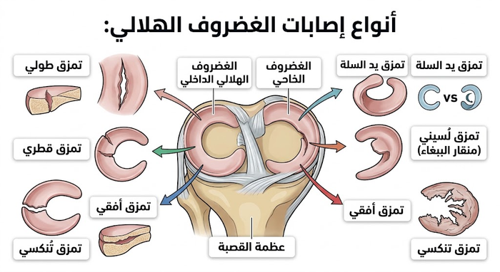
The most common type in young people and athletes; caused by a sudden forceful twisting of the knee under load. Common triggers include direction changes in football or basketball, a direct blow to the knee, or lifting heavy weights incorrectly. Tear patterns include longitudinal, radial, bucket-handle, horizontal, and complex tears.
Acute vs. Degenerative Tear: Side-by-Side Comparison
| Criterion | Acute (Traumatic) Tear | Degenerative Tear |
|---|---|---|
| Typical age group | 15–40 years | Over 40 years |
| Mechanism | Sudden twist / direct trauma | Progressive wear / trivial movement |
| Symptom onset | Sudden — within hours | Gradual — weeks to months |
| Repair (suture) candidacy | High (if in red zone) | Low — tissue quality poor |
| Conservative care success | Moderate — location-dependent | Good for mild symptoms |
| Association with osteoarthritis | Moderate — rises with neglect | High — often co-existing OA |
| Common patterns | Longitudinal, bucket-handle, radial | Horizontal, complex multi-directional |
| * Classification is for general awareness. Definitive type is established by clinical examination and MRI. | ||
Treatment Options
Conservative (Non-Surgical) Management
The first choice for small, partial, or degenerative tears that do not cause locking or instability. Key elements:
- R.I.C.E. protocol — Rest, Ice (15–20 min × several times/day), Compression, Elevation.
- NSAIDs to reduce pain and inflammation.
- Physiotherapy — progressive quadriceps and hamstring strengthening, range-of-motion, and proprioception exercises.
- Intra-articular injections — corticosteroid (acute inflammation) or PRP (biological healing stimulation).
Surgical Management (Arthroscopy)
Indicated when conservative care fails, or when mechanical symptoms (locking) are present. Three arthroscopic procedures:
- Meniscal Repair: suturing the torn edges — preferred whenever possible; preserves tissue and lowers long-term OA risk.
- Partial Meniscectomy: removal of the torn fragment only — most common procedure, especially for White Zone tears.
- Total Meniscectomy: avoided whenever possible; associated with substantially increased OA risk.

Comparing the Three Surgical Options
| Criterion | Repair (Suture) | Partial Meniscectomy | Total Meniscectomy |
|---|---|---|---|
| Goal | Preserve and heal the meniscus | Remove only the damaged fragment | Remove the entire meniscus |
| Ideal candidate | Young / athlete / red zone / acute tear | White zone / failed conservative / complex tear | Last resort only |
| Return to desk work | 2–4 weeks | 1–3 weeks | 2–4 weeks |
| Full return to sport | 4–6 months | 6–10 weeks (light activity) | 3–6 months |
| OA risk at 10 years | Lower — tissue preserved | Moderate — proportional to tissue removed | Very high |
| Pain-relief success rate | 75–95% | 80–90% | 70–85% (short-term) |
| Re-operation rate (5 years) | 10–20% | 5–15% | Higher — risk of eventual joint replacement |
| * Figures from AAOS, HSS, and Sihvonen et al. (NEJM 2013). Total meniscectomy is avoided in modern practice except in extreme cases. | |||
⚠ Clinical Note: Total meniscectomy is associated with a documented increase in early-onset knee osteoarthritis. Always discuss with your surgeon whether maximum meniscal tissue preservation is feasible in your case.
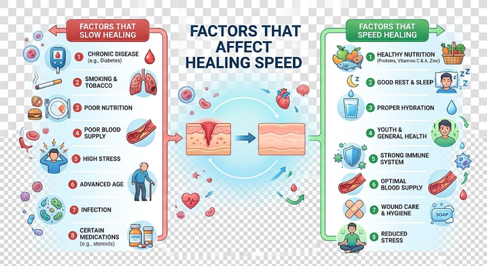
Factors That Affect Healing Speed
- Patient age: younger patients have superior regenerative capacity.
- Tear location: Red Zone tears heal far better than White Zone tears.
- Tear type and severity: simple longitudinal tears respond better than complex multi-pattern tears.
- General health: chronic disease or excess body weight slows recovery.
- Adherence to treatment: consistent physiotherapy attendance is decisive for outcome.
Recovery Timeline by Treatment Type
| Recovery Milestone | Conservative Care | Partial Meniscectomy | Meniscal Repair (Suture) |
|---|---|---|---|
| Acute pain relief | Week 1–2 | Day 3–7 post-op | Week 1–3 |
| Crutch-free walking | Usually not needed | Day 3–7 | Week 4–8 |
| Return to driving | Week 1–3 | Week 1–2 | Week 4–6 |
| Return to desk work | Week 1–2 | Week 1–3 | Week 2–4 |
| Normal walking | Week 2–4 | Week 2–4 | Week 6–10 |
| Low-impact sport (swimming, cycling) | Week 4–8 | Week 4–6 | Month 3–4 |
| Full competitive sport return | Month 3–6 | Week 6–10 | Month 4–6 |
| * Timelines are estimates and vary with physiotherapy adherence, patient age, and tear size. Competitive sport return requires functional testing and physician clearance. | |||
Frequently Asked Questions
Can a torn meniscus heal completely on its own without surgery?
Spontaneous biological healing depends entirely on local tissue vascularity:
- The meniscus has an uneven vascular distribution; only its outer peripheral rim receives direct arterial perfusion capable of generating a reparative fibrocartilage scar.
- Small, stable longitudinal tears located in this vascular zone can mend without surgery through rest, offloading, and structured rehabilitation.
- Tears in the avascular inner zone cannot physically seal together on their own, but conservative treatment can stabilize the joint, eliminate symptoms, and make the injury clinically silent.
How does the vascular anatomy (Red Zone vs. White Zone) determine healing potential?
The meniscus is divided into three distinct zones based on vascular supply:
- Red-Red Zone (Outer 10%–30%): Closest to the vascular synovial capsule, supplied by branches of the genicular arteries. Tears here have excellent healing capacity and can knit back together on their own.
- Red-White Zone (Middle 30%): A transitional area with limited microvascular penetration. Tears here heal inconsistently and often benefit from biological adjuvants like Platelet-Rich Plasma (PRP).
- White-White Zone (Inner 60%): Lacks blood vessels entirely and relies on passive nutrient diffusion from synovial fluid. Tears in this zone do not heal spontaneously or through surgical suturing.
What types of meniscus tears are most likely to heal without an operation?
Certain tear patterns have a favorable non-surgical prognosis:
- Small Longitudinal Tears: Stable vertical tears under 10 mm located within 3 to 4 mm of the meniscocapsular junction.
- Partial-Thickness Tears: Tears that do not penetrate completely through both the superior and inferior articular surfaces of the fibrocartilage.
- Degenerative Tears in Patients Over 40: Gradual wear tears that are not locked or displaced; clinical trials show physical therapy produces functional outcomes equivalent to partial meniscectomy for these tears.
What non-surgical treatments encourage natural meniscus recovery?
A structured conservative protocol provides the optimal mechanical and biological environment for healing:
- Protection & Optimal Loading (POLICE): Relative rest during acute flares while avoiding prolonged joint immobilization to maintain chondrocyte nutrition.
- Targeted Physical Rehabilitation: Strengthening the vastus medialis, hamstrings, and hip abductors builds a muscular shock absorber that relieves compressive joint loads by up to 30% to 40%.
- Sensory Proprioceptive Retraining: Balance drills on foam pads or wobble boards eliminate micro-shearing during walking.
- External Support: Compressive knee sleeves or unloader braces minimize lateral and rotational stress across the damaged compartment.
How do biological injections like PRP help a torn meniscus heal?
Platelet-Rich Plasma (PRP) delivers autologous signaling proteins to augment natural repair:
- Growth Factor Delivery: High concentrations of TGF-β, PDGF, and VEGF stimulate local cellular migration, extracellular matrix deposition, and micro-angiogenesis.
- Anti-Inflammatory Modulation: PRP suppresses degradative synovial cytokines (such as IL-1β and TNF-α), reducing chronic swelling and protecting the joint’s hyaline cartilage.
- Ultrasound Targeting: Point-of-care musculoskeletal ultrasound guidance ensures the biologic concentrate is deposited directly into the perimeniscal tissue and tear margins.
How long does it take for a meniscus tear to heal on its own?
A successful non-operative recovery spans 2 to 4 months through specific phases:
- Phase 1 (Weeks 1 to 3): Inflammation, joint effusion, and resting pain subside through relative offloading, cryotherapy, and isometric muscle firing.
- Phase 2 (Weeks 4 to 8): Fibrocartilaginous scar tissue forms in vascular tears, dynamic range of motion normalizes, and low-impact stationary cycling begins.
- Phase 3 (Weeks 8 to 16): Collagen bridges remodel and mature, allowing full unassisted ambulation, functional strength recovery, and a gradual return to active recreational tasks.
What clinical signs indicate that a meniscus tear is successfully healing?
Key objective indicators confirming that the joint is stabilizing non-operatively include:
- Absence of Effusion: The knee stays cool and dry without swelling after walking or low-impact workouts.
- Clear Joint Line: Focal palpation along the medial or lateral tibiofemoral joint line no longer elicits sharp local pain.
- Full Terminal Extension: The knee straightens completely (0 degrees) without tightness or catching in the posterior capsule.
- Negative Provocative Tests: Passive flexion with internal/external tibial rotation (McMurray test) produces no pain, clicking, or subluxation sensations.
What are the common daily mistakes that prevent a meniscus tear from healing?
Certain physical activities create destructive joint forces that disrupt repair:
- Deep Knee Flexion Past 90 Degrees: Deep squats, kneeling, and floor sitting concentrate high compressive forces on the posterior horns, expanding the tear gap.
- Sudden Pivoting: Twisting the torso with a firmly planted foot generates severe shear forces across fragile healing fibers.
- Premature Impact Loading: Returning to running on pavement, jumping, or sports before quadriceps symmetry reaches at least 85%–90%.
- Complete Inactivity: Avoiding movement entirely leads to rapid quadriceps atrophy and starves the joint cartilage of synovial fluid circulation.
What happens if an avascular (White Zone) tear does not heal?
An unhealed inner tear follows one of two clinical paths based on mechanical stability:
- Stable & Asymptomatic: If the tear edges stay stable and do not catch, strong periarticular muscles can support the joint, allowing the patient to live normally without surgery.
- Unstable & Chondrodestructive: If loose fragments flip into the joint space, they cause persistent swelling and act like an abrasive wedge against the femoral and tibial surfaces. Over time, this accelerates articular cartilage breakdown and leads to premature unicompartmental osteoarthritis, which warrants arthroscopic trimming (partial meniscectomy).
When does natural healing fail, requiring arthroscopic surgery?
Specific structural indicators signal that conservative management is no longer appropriate:
- Mechanical Joint Locking: A displaced bucket-handle fragment physically blocks full knee extension; forced motion risks permanently damaging the tissue, making urgent arthroscopic reduction necessary.
- Meniscal Root Avulsions: Tears at the anterior or posterior bone insertions cause the meniscus to extrude outward, completely eliminating its load-bearing capacity and mimicking a total meniscectomy.
- Refractory Symptoms: Ongoing, sharp, disabling pain, recurrent effusions, or functional instability that fails to improve after 8 to 12 weeks of compliant physical rehabilitation.
International References
- AAOS OrthoInfo. Meniscus Tears. American Academy of Orthopaedic Surgeons. 2023. Available at: orthoinfo.aaos.org
- Mayo Clinic Staff. Torn meniscus – Symptoms and causes. Mayo Clinic. 2024. Available at: mayoclinic.org
- Hospital for Special Surgery (HSS). Meniscus Tears: Causes, Symptoms, and Treatment Options. 2023. Available at: hss.edu
- Cleveland Clinic. Meniscus Tear. 2023. Available at: my.clevelandclinic.org
- Katz JN, et al. Surgery versus Physical Therapy for a Meniscal Tear and Osteoarthritis. New England Journal of Medicine. 2013;368(18):1675–1684. DOI: 10.1056/NEJMoa1301408
- Sihvonen R, et al. Arthroscopic Partial Meniscectomy versus Sham Surgery for a Degenerative Meniscal Tear (FIDELITY). New England Journal of Medicine. 2013;369:2515–2524. DOI: 10.1056/NEJMoa1305189
- Thorlund JB, et al. Arthroscopic surgery for degenerative knee: systematic review and meta-analysis of benefits and harms. BMJ. 2015;350:h2747. DOI: 10.1136/bmj.h2747
- Lohmander LS, et al. The long-term consequence of ACL and meniscus injuries: osteoarthritis. American Journal of Sports Medicine. 2007;35(10):1756–1769. DOI: 10.1177/0363546507307396