
Quick answer
A PET-CT scan for sarcoma combines an image of cell activity (PET) with a CT scan that pins down location. A radioactive sugar (FDG) is injected, and active cells, including cancer cells, take up more of it and light up. In bone and soft tissue sarcoma we use it to stage some malignant tumours and to check response to treatment; most benign tumours do not need it.
✅ Last medically reviewed by Prof. Dr. Ibrahim Shaarawi, Professor of Orthopaedic Surgery, on 27 October 2026
Some patients ask for a PET-CT at the start, thinking it is “the most complete test”. It is very useful in specific situations and can muddy the picture in others. This guide covers when it helps, when we skip it, and how to prepare. It is part of our bone and soft tissue tumor surgery guide.
How PET-CT works
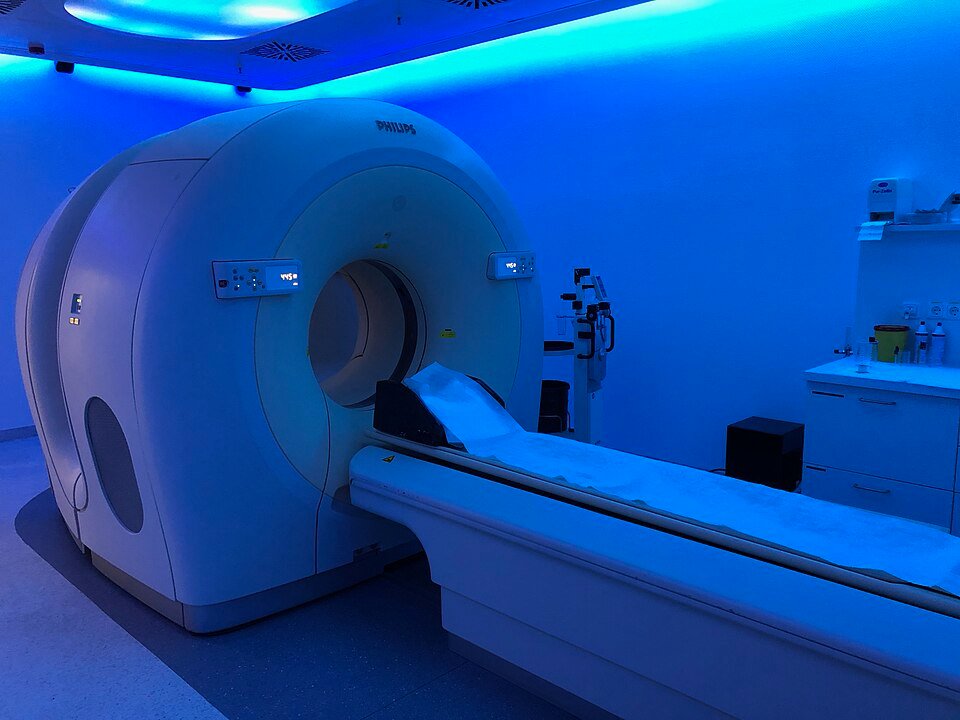
Rapidly dividing cells burn more sugar. Sugar tagged with a radioactive label collects in those cells and is picked up by the PET scanner, while the CT taken in the same session shows exactly where that activity sits: bone, lung or lymph node. The report gives a value called SUV for how intense the uptake is.
When we order it for bone and soft tissue tumours
- Staging Ewing sarcoma and some high-grade sarcomas, to look for spread to bone and lymph nodes (Seth et al., 2022).
- Selected patients with osteosarcoma or soft tissue sarcoma, as guidelines allow (ESMO, 2021).
- Multiple myeloma, to map disease and assess response (IMWG, 2019).
- Checking response to chemotherapy, or telling recurrence from scar tissue after surgery.
When we skip it, and when it can mislead
- Benign tumours: most do not need it. Some, such as giant cell tumor of bone and PVNS, can light up and cause needless worry.
- Inflammation, healing fractures and recent surgery: all take up sugar and glow.
- The lungs: it does not replace a chest CT for small lung nodules.
- The main tumour: it does not replace MRI for bone tumors for defining the tumour’s edges before surgery.
Video: Important steps after a tumour is diagnosed
Key numbers to know
- In Ewing sarcoma, PET-CT detected bone metastases with 83.9% sensitivity and 93.2% specificity, but its sensitivity for lung metastases was only 76.1% (Seth et al., 2022).
- In a meta-analysis of 67 studies on detecting bone metastases, per-patient sensitivity was 90.6% for MRI, 89.7% for PET and 86% for bone scan, with specificity of 95.4%, 96.8% and 81.4% respectively (Yang et al., 2011).
- The PET-CT tracer needs 30 to 60 minutes to spread through the body, then scanning takes about 30 minutes: under two minutes for the CT and 20 to 30 minutes for the PET images (RadiologyInfo).
| Test | What it shows well | When we order it |
|---|---|---|
| X-ray | Bone shape, tumour edges and calcification | Always the first test for a bone lesion |
| MRI | Spread inside the marrow and into muscle, nerves and vessels | Before biopsy and surgery, and for any soft tissue mass |
| CT scan | Bone cortex, fine calcification and the lungs | Surgical planning, and chest CT for staging malignant tumours |
| Bone scan | Active spots across the whole skeleton | Looking for more than one bone lesion |
| PET-CT | Tumour activity in the whole body | Staging some malignant tumours and checking treatment response |
At Prof. Dr. Ibrahim Shaarawi’s clinics in Nasr City, Mohandeseen and New Cairo (5th Settlement) we look at the images themselves, not just the report, and tell you whether another test is needed or whether what you have is enough to decide.
Preparing for the scan
- Fast for several hours as instructed by the centre and avoid sugary drinks. Water is fine.
- People with diabetes get special instructions for insulin and tablets, because high blood sugar spoils the images.
- Avoid hard exercise the day before, as tired muscles soak up sugar.
- After the injection you rest quietly for 30 to 60 minutes without much talking or moving.
- Tell the team if you are pregnant or breastfeeding, and ask about storing milk in advance.
Understanding the result
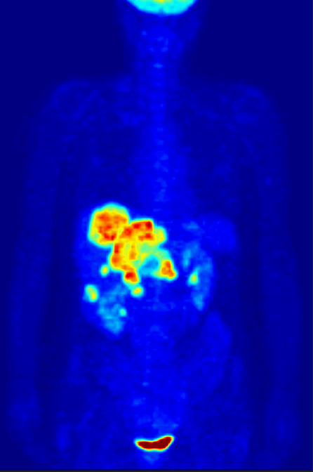
The brain, heart, kidneys and bladder light up in everyone, and that is normal. An SUV number is not a diagnosis on its own; we read it alongside MRI, CT and the patient’s history. If an unexpected spot appears, the next step is usually a targeted scan or a biopsy, not worry over a number in the report.
Radiation from PET-CT
The tracer dose is small and decays quickly, and the CT adds its own dose (RadiologyInfo). The test is safe when there is a real reason for it, but we do not repeat it without one, and we choose between it and a bone scan for bone tumors according to the tumour type.
Frequently asked questions about the PET-CT scan for sarcoma
Does PET-CT detect every type of cancer?
No. Some tumours take up little sugar, and some benign conditions and inflammation take up a lot, so results are read with the other tests.
Do I need PET-CT for a benign bone tumour?
Usually not. X-ray and MRI are enough for most benign tumours, and some can light up on PET without being malignant.
How long should I fast before the scan?
Several hours as the centre instructs, avoiding sugar and sweet drinks; water is allowed.
How long does the scan take?
A 30 to 60 minute wait after the injection, then about 30 minutes of scanning.
Does PET-CT replace a chest CT?
No. A dedicated chest CT is better at finding small lung nodules, so we order it alongside PET in malignant tumours.
To have your scans reviewed by a professor of orthopaedic oncology, book a consultation, call +20 155 240 4488 or message us on WhatsApp at +20 155 554 7181. Bring the images on CD, not only the report.
Medically reviewed by Prof. Dr. Ibrahim Shaarawi, Professor of Orthopaedic Surgery. This article is general medical education and does not replace an examination or the plan your own doctor sets for you. See our medical disclaimer.
Related bone and soft tissue tumor guides
Related tests and guides for diagnosing bone tumours:
- Bone and soft tissue tumor surgery
- MRI for bone tumors
- Bone scan for bone tumors
- Bone cancer symptoms
- Giant cell tumor of bone
References
- Seth N, et al. (18) F-FDG PET and PET/CT as a diagnostic method for Ewing sarcoma: A systematic review and meta-analysis. Pediatr Blood Cancer. 2022;69(3):e29415.
- Gronchi A, et al. Soft tissue and visceral sarcomas: ESMO-EURACAN-GENTURIS Clinical Practice Guidelines for diagnosis, treatment and follow-up(☆). Ann Oncol. 2021;32(11):1348-1365.
- Hillengass J, et al. International myeloma working group consensus recommendations on imaging in monoclonal plasma cell disorders. Lancet Oncol. 2019;20(6):e302-e312.
- RadiologyInfo.org (ACR and RSNA). Radiation Dose in X-Ray and CT Exams.
- Yang HL, et al. Diagnosis of bone metastases: a meta-analysis comparing ¹⁸FDG PET, CT, MRI and bone scintigraphy. Eur Radiol. 2011;21(12):2604-17.
- RadiologyInfo.org (ACR and RSNA). Positron Emission Tomography and Computed Tomography (PET/CT).