
Quick answer
A bone scan for bone tumors is a nuclear medicine test: a small amount of radioactive tracer is injected into a vein, and two to four hours later the whole skeleton is imaged, with active areas showing as dark “hot spots”. Its main use in bone tumours is to look for other lesions anywhere in the skeleton. It cannot say what a hot spot is: fractures, inflammation, arthritis and benign tumours all light up too.
✅ Last medically reviewed by Prof. Dr. Ibrahim Shaarawi, Professor of Orthopaedic Surgery, on 27 October 2026
Many people are frightened by the word “nuclear” and by a report mentioning an “area of increased uptake”. This guide explains how the scan works, when we really need it, what a hot spot means, and when another test is better. It is part of our bone and soft tissue tumor surgery guide.
How a bone scan works
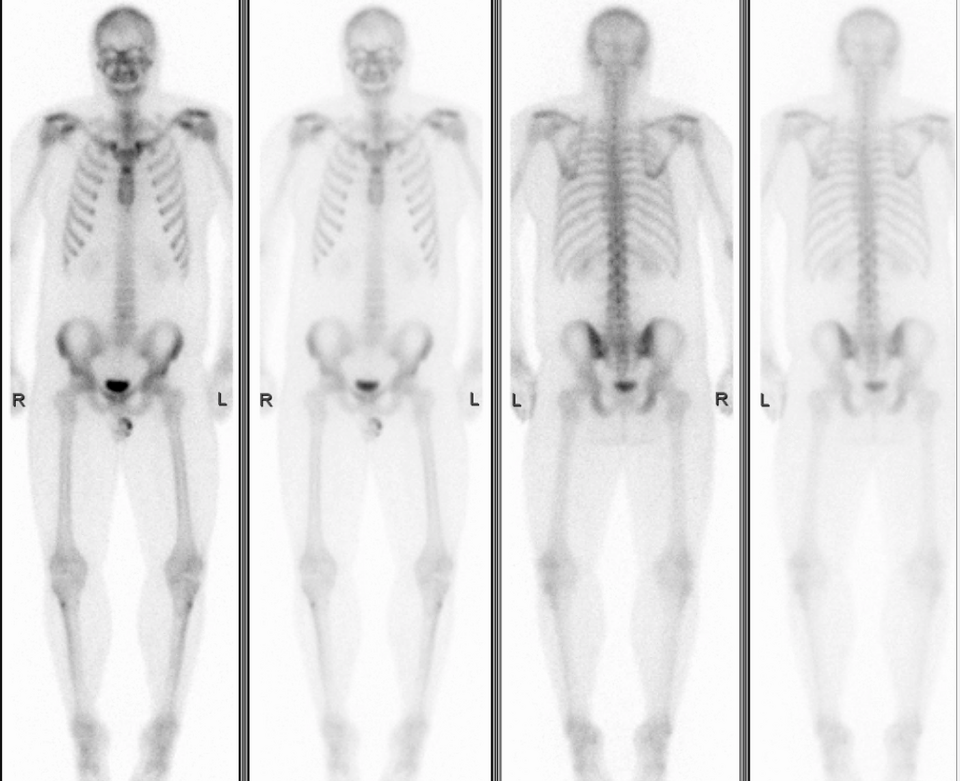
The tracer, usually technetium attached to a phosphate compound, sticks to bone where new bone is being laid down quickly (SNMMI procedure standard, 2018). Any area of increased activity, whether a tumour, a healing fracture or inflammation, takes up more and looks darker. The strength of the test is that it images the whole body at once; the weakness is lower image detail than MRI or CT.
When we order it for bone tumours
- Staging primary malignant bone tumours such as osteosarcoma, to look for other skeletal lesions (ESMO, 2021).
- Looking for cancer that has spread to bone from another organ, such as breast or prostate.
- Bone pain with a normal X-ray, as in stress fractures.
- Finding out whether a tumour is single or one of several.
What a hot spot means
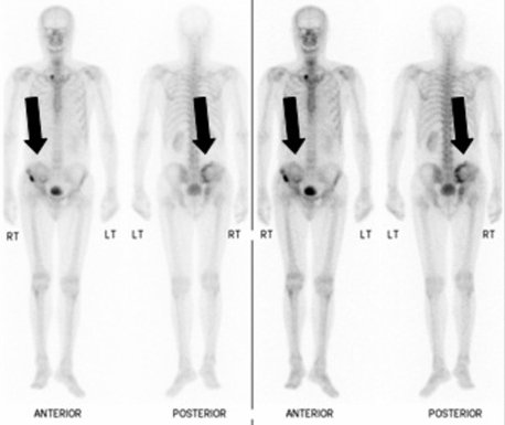
A hot spot only means the bone is active there. It does not say why. Common causes:
- An old or recent fracture, including stress fractures.
- Arthritis of joints and the spine.
- Inflammation or infection of bone.
- Benign tumours, including enchondroma, which is usually active on bone scan (OrthoInfo).
- Malignant tumours or metastases.
So we usually follow up with MRI for bone tumors or CT of the spot, and sometimes a biopsy (RadiologyInfo).
Video: Important steps after a tumour is diagnosed
Key numbers to know
- In a meta-analysis of 67 studies on detecting bone metastases, per-patient sensitivity was 90.6% for MRI, 89.7% for PET and 86% for bone scan, with specificity of 95.4%, 96.8% and 81.4% respectively (Yang et al., 2011).
- After the tracer injection the wait is usually two to four hours, patients are asked to drink four to six glasses of water, and breastfeeding mothers use formula for one to two days (RadiologyInfo).
| Test | What it shows well | When we order it |
|---|---|---|
| X-ray | Bone shape, tumour edges and calcification | Always the first test for a bone lesion |
| MRI | Spread inside the marrow and into muscle, nerves and vessels | Before biopsy and surgery, and for any soft tissue mass |
| CT scan | Bone cortex, fine calcification and the lungs | Surgical planning, and chest CT for staging malignant tumours |
| Bone scan | Active spots across the whole skeleton | Looking for more than one bone lesion |
| PET-CT | Tumour activity in the whole body | Staging some malignant tumours and checking treatment response |
At Prof. Dr. Ibrahim Shaarawi’s clinics in Nasr City, Mohandeseen and New Cairo (5th Settlement) we look at the images themselves, not just the report, and tell you whether another test is needed or whether what you have is enough to decide.
When a bone scan is not the right test
- Multiple myeloma: it punches holes in bone without new bone formation, so it may not show. The IMWG recommends whole-body low-dose CT, PET-CT or MRI instead (Hillengass et al., 2019).
- Mapping the main tumour: MRI is the right test, not a bone scan.
- Some sarcomas: such as Ewing sarcoma, where a PET-CT scan for sarcoma may be preferred for staging.
Steps and preparation
- No fasting is usually needed.
- Tell the team if you took a bismuth medicine or had a barium X-ray within four days, as these interfere with the images.
- Tell them if you are pregnant or breastfeeding.
- After the injection you wait two to four hours, drink plenty of water, then empty your bladder just before imaging.
- The scan is painless, you can resume normal activity afterwards, and the small radiation dose leaves in the urine within a short time.
Frequently asked questions about the bone scan for bone tumors
Does a hot spot on a bone scan mean cancer?
No. It only means bone activity, which can come from a fracture, arthritis, inflammation or a benign tumour; MRI or CT usually follows.
Is a bone scan dangerous?
The radiation dose is small and leaves the body quickly in the urine, and you are no risk to people around you after the test.
How long does it take?
Two to four hours of waiting after the injection, then the imaging itself; the visit can take a few hours in total.
Can a breastfeeding mother have a bone scan?
Yes, using formula for one to two days afterwards and discarding breast milk during that time.
Which is better, bone scan or PET-CT?
It depends on the tumour. A bone scan suits the search for bone lesions in many tumours; PET-CT is more accurate in some, such as Ewing sarcoma and myeloma.
To have your scans reviewed by a professor of orthopaedic oncology, book a consultation, call +20 155 240 4488 or message us on WhatsApp at +20 155 554 7181. Bring the images on CD, not only the report.
Medically reviewed by Prof. Dr. Ibrahim Shaarawi, Professor of Orthopaedic Surgery. This article is general medical education and does not replace an examination or the plan your own doctor sets for you. See our medical disclaimer.
Related bone and soft tissue tumor guides
Related tests and guides for diagnosing bone tumours:
- Bone and soft tissue tumor surgery
- MRI for bone tumors
- PET-CT scan for sarcoma
- Enchondroma
- Bone cancer symptoms
References
- Bartel TB, et al. SNMMI Procedure Standard for Bone Scintigraphy 4.0. J Nucl Med Technol. 2018;46(4):398-404.
- Strauss SJ, et al. Bone sarcomas: ESMO-EURACAN-GENTURIS-ERN PaedCan Clinical Practice Guideline for diagnosis, treatment and follow-up. Ann Oncol. 2021;32(12):1520-1536.
- American Academy of Orthopaedic Surgeons (OrthoInfo). Enchondroma.
- Hillengass J, et al. International myeloma working group consensus recommendations on imaging in monoclonal plasma cell disorders. Lancet Oncol. 2019;20(6):e302-e312.
- Yang HL, et al. Diagnosis of bone metastases: a meta-analysis comparing ¹⁸FDG PET, CT, MRI and bone scintigraphy. Eur Radiol. 2011;21(12):2604-17.
- RadiologyInfo.org (ACR and RSNA). Bone Scan (Bone Scintigraphy).