
Quick answer
MRI for bone tumors shows how far a tumour extends inside the bone and into muscle, nerves and blood vessels, so we order it before a biopsy and before any surgery. It uses no ionising radiation and usually takes 15 minutes to an hour. A plain X-ray is still the first test for any bone lesion; MRI adds to it rather than replacing it.
✅ Last medically reviewed by Prof. Dr. Ibrahim Shaarawi, Professor of Orthopaedic Surgery, on 27 October 2026
A question we hear often: “I’ve had an MRI, do I need anything else?” The answer depends on what the MRI showed and on the type of tumour. This guide covers what MRI shows, why it must come before a biopsy, and how to read the common report terms. It is part of our bone and soft tissue tumor surgery guide.
Why MRI follows the X-ray
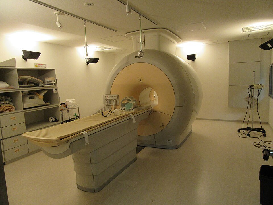
The X-ray shows the shape and edges of a lesion, which tells us whether it looks quiet or aggressive. If the X-ray is inconclusive or the lesion looks aggressive, MRI or CT is the next step to define its extent and plan the biopsy and surgery, according to the American College of Radiology (ACR, 2025). For a soft tissue mass that cannot be confirmed as benign on examination, imaging is needed before any intervention (ACR, 2023).
What MRI shows in bone and soft tissue tumours
- Spread within the marrow and any other lesions in the same bone, which is why the whole bone and both neighbouring joints are imaged in malignant tumours (ESMO, 2021).
- The size of any part that has broken out of the bone into muscle.
- How close the tumour is to the main nerves, vessels and joint, which decides whether the limb can be saved.
- Tissue type: fat in lipomas, fluid in cysts, cartilage in an enchondroma, haemosiderin in pigmented villonodular synovitis.
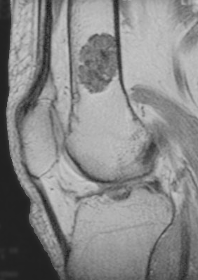
Why MRI must come before a biopsy
A biopsy causes bleeding and swelling around the tumour, which changes how it looks on MRI and blurs its edges. Just as important, the needle or incision track has to lie where the surgeon will later remove it with the tumour. So we get the MRI first, then the tumour surgeon plans the biopsy route.
Video: Not every lump or mass in the body is cancer
Key numbers to know
- Cartilage tumours were found by chance on knee MRI in 2.8% of 1,285 people, with a mean size of 12 mm (Stomp et al., 2015). In a review of 44,762 knee MRIs the rate was 1.45%, and 72.9% sat in the lower femur (Woltsche et al., 2023).
- In a meta-analysis of 67 studies on detecting bone metastases, per-patient sensitivity was 90.6% for MRI, 89.7% for PET and 86% for bone scan, with specificity of 95.4%, 96.8% and 81.4% respectively (Yang et al., 2011).
- An MRI usually takes 15 minutes to an hour, and after contrast injection patients wait up to 30 minutes to check for a reaction (NHS).
| Test | What it shows well | When we order it |
|---|---|---|
| X-ray | Bone shape, tumour edges and calcification | Always the first test for a bone lesion |
| MRI | Spread inside the marrow and into muscle, nerves and vessels | Before biopsy and surgery, and for any soft tissue mass |
| CT scan | Bone cortex, fine calcification and the lungs | Surgical planning, and chest CT for staging malignant tumours |
| Bone scan | Active spots across the whole skeleton | Looking for more than one bone lesion |
| PET-CT | Tumour activity in the whole body | Staging some malignant tumours and checking treatment response |
At Prof. Dr. Ibrahim Shaarawi’s clinics in Nasr City, Mohandeseen and New Cairo (5th Settlement) we look at the images themselves, not just the report, and tell you whether another test is needed or whether what you have is enough to decide.
Contrast and preparing for the scan
- Contrast (gadolinium): given into a vein for most tumour scans, because it separates living solid tumour from dead or fluid areas. Kidney function is checked when needed.
- Metal in the body: tell the team about a pacemaker, plates, screws or metal fragments.
- Claustrophobia: a mild sedative can be given; do not drive for 24 hours afterwards.
- Noise: the scanner makes loud knocking sounds, and earplugs are provided.
Pregnancy is not an absolute bar to MRI without contrast, but tell your doctor; contrast is avoided in pregnancy unless essential.
How to read your MRI report
Reports use terms that can sound alarming. The common ones:
- Low / high signal on T1, T2: describes how the tissue looks on different sequences; on its own it does not mean benign or malignant.
- Enhancement: the tissue takes up contrast, meaning it is alive and has a blood supply.
- Cortical breach / destruction: a break in the bone’s outer shell, worth prompt review.
- Soft tissue component: part of the tumour has left the bone.
- Incidental finding: something found by chance, usually unrelated to why the scan was done.
If your knee MRI was done for an injury, see also reading a knee MRI report.
When MRI alone is not enough
MRI only covers the area scanned. In malignant tumours we also need a chest CT, because the lungs are the commonest site of spread from bone and soft tissue sarcomas (ACR, 2022), and a whole-body test such as a bone scan for bone tumors or PET-CT scan for sarcoma depending on the tumour. CT also shows the bone cortex and fine calcification more clearly than MRI.
Frequently asked questions about MRI for bone tumors
Can MRI tell whether a tumour is malignant?
It gives strong clues, but for unclear lesions the final diagnosis comes from a biopsy taken after the MRI.
Do I need MRI with contrast?
For most tumour scans, yes, because contrast shows the living part of the tumour and helps plan the biopsy and surgery.
Does MRI use radiation?
No. MRI uses a magnetic field and radio waves, not X-rays.
Can I have an MRI with metal plates and screws?
Most modern implants are MRI-safe, though they can distort the image nearby. Always tell the team before the scan.
Why did my doctor ask for a new MRI when I already have one?
The earlier scan may not have covered the whole bone, may lack contrast, may be old, or may have been done after a biopsy.
To have your scans reviewed by a professor of orthopaedic oncology, book a consultation, call +20 155 240 4488 or message us on WhatsApp at +20 155 554 7181. Bring the images on CD, not only the report.
Medically reviewed by Prof. Dr. Ibrahim Shaarawi, Professor of Orthopaedic Surgery. This article is general medical education and does not replace an examination or the plan your own doctor sets for you. See our medical disclaimer.
Related bone and soft tissue tumor guides
Related tests and guides for diagnosing bone tumours:
- Bone and soft tissue tumor surgery
- Bone scan for bone tumors
- PET-CT scan for sarcoma
- Enchondroma
- Reading a knee MRI report
References
- Ahlawat S, et al. ACR Appropriateness Criteria® Suspected Primary Bone Tumors: 2024 Update. J Am Coll Radiol. 2025;22(5S):S440-S454.
- Garner HW, et al. ACR Appropriateness Criteria® Soft Tissue Masses: 2022 Update. J Am Coll Radiol. 2023;20(5S):S234-S245.
- Strauss SJ, et al. Bone sarcomas: ESMO-EURACAN-GENTURIS-ERN PaedCan Clinical Practice Guideline for diagnosis, treatment and follow-up. Ann Oncol. 2021;32(12):1520-1536.
- Stanborough R, et al. ACR Appropriateness Criteria® Malignant or Aggressive Primary Musculoskeletal Tumor-Staging and Surveillance: 2022 Update. J Am Coll Radiol. 2022;19(11S):S374-S389.
- Stomp W, et al. Prevalence of cartilaginous tumours as an incidental finding on MRI of the knee. Eur Radiol. 2015;25(12):3480-7.
- Yang HL, et al. Diagnosis of bone metastases: a meta-analysis comparing ¹⁸FDG PET, CT, MRI and bone scintigraphy. Eur Radiol. 2011;21(12):2604-17.
- NHS. MRI scan.
- Woltsche JN, et al. Prevalence and characteristics of benign cartilaginous tumours of the knee joint as identified on MRI scans. Cancer Imaging. 2023;23(1):50.