
Quick answer
Pigmented villonodular synovitis (PVNS) is a benign tumour of the joint lining, and about 80% of cases involve the knee. It does not spread to other organs, but left for years it can damage cartilage and bone. MRI makes the diagnosis, and treatment means removing the diseased lining, arthroscopically or through open surgery depending on the type.
✅ Last medically reviewed by Prof. Dr. Ibrahim Shaarawi, Professor of Orthopaedic Surgery, on 27 October 2026
Pathologists now call it tenosynovial giant cell tumour (TGCT), though many reports still say PVNS. Patients often reach us after months of knee swelling that was treated as arthritis or a cartilage tear, until an MRI showed the real cause. This guide explains the two types and why each is treated differently. It is part of our bone and soft tissue tumor surgery guide.
What is pigmented villonodular synovitis?
The synovium is the thin lining that produces joint fluid. In PVNS its cells multiply, so the lining thickens into fronds and nodules. Small repeated bleeds leave a brown iron pigment called haemosiderin in the tissue, which is where the word “pigmented” comes from.
- Localized type: one or a few nodules in part of the joint. Easier to treat.
- Diffuse type: involves most of the lining and can extend behind the knee or into nearby muscle. Harder to clear and more likely to come back.
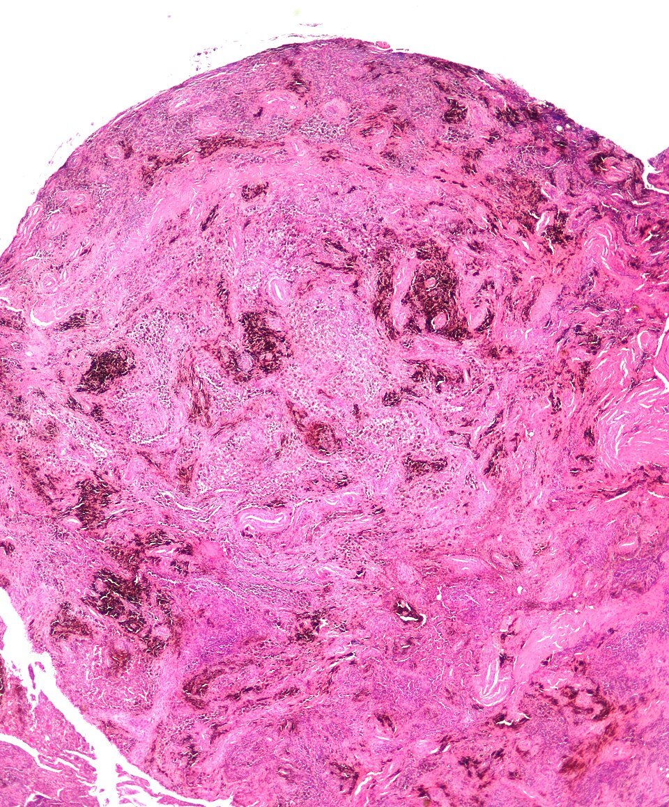
It is benign in the strict sense: an Italian series of 294 patients recorded no spread to other organs (Palmerini et al., 2015). The problem is local recurrence after surgery, mainly in the diffuse type.
Symptoms that should raise suspicion
- Knee swelling that keeps coming back without a clear injury.
- Fluid drained from the knee that is red or brown instead of clear yellow.
- Pain and stiffness that build up over months or years.
- Sudden locking or catching, more typical of a localized nodule.
- A sense of fullness or a lump behind the knee.
It usually appears between 20 and 50, is slightly more common in women, and normally affects one joint (AAOS OrthoInfo). Because it mimics a meniscus tear, arthritis or a Baker’s cyst, it is easy to miss without an MRI.
Diagnosis: why MRI matters most
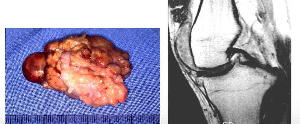
X-rays are often normal early on; later they may show erosions on both sides of the joint, particularly in the hip. MRI shows a thickened lining with dark areas from haemosiderin, and these areas look even darker on gradient-echo sequences, a sign that is close to diagnostic (Murphey et al., 2008).
MRI also maps the disease: front of the knee only, behind it as well, or outside the joint capsule. That map decides the operation. Our guide to MRI for bone tumors explains the report. A biopsy is occasionally needed when the picture is unclear.
Key numbers to know
- In a nationwide Dutch study of 4,503 pathology reports, the knee was the most affected joint (46% of localized and 64% of diffuse cases), and 23% of diffuse cases needed another operation for recurrence versus 9% of localized ones (Mastboom et al., 2017).
- In pooled data on 630 knees, PVNS came back in 21.8%; in diffuse disease, open or combined open-plus-arthroscopic synovectomy had fewer recurrences than arthroscopy alone (Mollon et al., 2015).
- In the largest international series of diffuse-type disease (966 operated patients), 44% had a recurrence and 55% were recurrence-free at 5 years (Mastboom et al., 2019).
- In the ENLIVEN trial, the targeted drug pexidartinib shrank the tumour in 39% of patients versus 0% on placebo, with liver toxicity that needs monitoring (Tap et al., The Lancet 2019). In the MOTION trial, vimseltinib gave a 40% response versus 0% (Gelderblom et al., The Lancet 2024).
| Treatment | When we choose it | What to expect |
|---|---|---|
| MRI surveillance | Small tumour without symptoms, or a patient not ready for surgery | MRI every 6 to 12 months to watch the size |
| Arthroscopic excision | Localized (single nodule) disease and front-of-knee diffuse disease | Small incisions and quick return to walking; localized disease rarely returns |
| Open or combined open and arthroscopic synovectomy | Diffuse disease, especially behind the knee | Better disease control with longer rehabilitation |
| Targeted drugs (CSF1R inhibitors) | Recurrent disease or tumour that cannot be removed without major harm | Shrinkage in about 4 in 10 patients, with blood test monitoring |
| Joint replacement | Advanced cartilage damage after years of disease | Pain relief, with synovectomy in the same operation |
At Prof. Dr. Ibrahim Shaarawi’s clinics in Nasr City, Mohandeseen and New Cairo (5th Settlement) we look at the images themselves, not just the report, explain what they show, and agree with you on follow-up or the right operation.
Localized versus diffuse: why treatment differs
A localized nodule can be removed whole with a margin of lining, and it seldom returns. AAOS quotes five-year recurrence of 10% to 30% for localized disease and 20% to 70% for diffuse disease (OrthoInfo). The gap exists because diffuse disease spreads into recesses that are hard to reach, and any lining left behind can regrow.
Video: Arthroscopic removal of a synovial tumour (PVNS) of the knee
Surgery: arthroscopic or open?
- Arthroscopy: suits localized disease and diffuse disease in the front of the knee. Small incisions, and most people walk the same or the next day.
- Open or combined open and arthroscopic synovectomy: for diffuse disease, especially behind the knee. Pooled data show fewer recurrences than arthroscopy alone (Mollon et al., 2015).
- Joint replacement: when years of disease have worn the cartilage away; synovectomy and total knee replacement are done together.
We plan the operation from the MRI and send all removed tissue to the lab. Physiotherapy starts early to regain knee bend. To read about the technique itself, see knee arthroscopy.
Targeted drugs and radiotherapy
Oral drugs that block the CSF1R signal the tumour depends on, such as pexidartinib and vimseltinib, are now available. We consider them when disease keeps returning, or when complete removal would badly damage the joint. They do not replace surgery for most patients and need liver and blood monitoring. Radiotherapy has a limited role today, with weak evidence, and is kept for selected recurrent cases.
Follow-up after surgery
Localized disease usually needs only short follow-up. Diffuse disease is followed for years, typically with MRI every 6 to 12 months at first (OrthoInfo). New swelling or fresh blood in the joint is a reason to repeat the MRI early.
Frequently asked questions about pigmented villonodular synovitis
Is pigmented villonodular synovitis cancer?
No. It is a benign tumour that does not spread to the lungs or other organs, but it can damage the joint and may return after surgery.
Does PVNS come back after surgery?
Recurrence is uncommon in the localized type and much more common in the diffuse type, which is why diffuse disease is followed with MRI for years.
How soon can I walk after arthroscopic removal?
Most patients walk with support the same or the next day, then physiotherapy restores movement over the first weeks.
Can it be treated with injections or tablets alone?
Injections do not treat it. Targeted tablets are an option for recurrent or inoperable disease but do not replace surgery for most patients.
Can it affect both knees?
Very rarely. It almost always involves a single joint, most often the knee, then the hip and ankle.
To have your scans reviewed by a professor of orthopaedic oncology, book a consultation, call +20 155 240 4488 or message us on WhatsApp at +20 155 554 7181. Bring the images on CD, not only the report.
Medically reviewed by Prof. Dr. Ibrahim Shaarawi, Professor of Orthopaedic Surgery. This article is general medical education and does not replace an examination or the plan your own doctor sets for you. See our medical disclaimer.
Related bone and soft tissue tumor guides
Related guides on this condition and its treatment:
- Bone and soft tissue tumor surgery
- Synovial chondromatosis
- MRI for bone tumors
- Knee arthroscopy
- Baker’s cyst
- Total knee replacement
References
- American Academy of Orthopaedic Surgeons (OrthoInfo). Tenosynovial Giant Cell Tumor (Pigmented Villonodular Synovitis).
- Murphey MD, et al. Pigmented villonodular synovitis: radiologic-pathologic correlation. Radiographics. 2008;28(5):1493-518.
- Palmerini E, et al. Tenosynovial giant cell tumour/pigmented villonodular synovitis: outcome of 294 patients before the era of kinase inhibitors. Eur J Cancer. 2015;51(2):210-7.
- Mollon B, et al. The effect of surgical synovectomy and radiotherapy on the rate of recurrence of pigmented villonodular synovitis of the knee: an individual patient meta-analysis. Bone Joint J. 2015;97-B(4):550-7.
- Mastboom MJL, et al. Higher incidence rates than previously known in tenosynovial giant cell tumors. Acta Orthop. 2017;88(6):688-694.
- Mastboom MJL, et al. Surgical outcomes of patients with diffuse-type tenosynovial giant-cell tumours: an international, retrospective, cohort study. Lancet Oncol. 2019;20(6):877-886.
- Tap WD, et al. Pexidartinib versus placebo for advanced tenosynovial giant cell tumour (ENLIVEN): a randomised phase 3 trial. Lancet. 2019;394(10197):478-487.
- Gelderblom H, et al. Vimseltinib versus placebo for tenosynovial giant cell tumour (MOTION): a multicentre, randomised, double-blind, placebo-controlled, phase 3 trial. Lancet. 2024;403(10445):2709-2719.