
Giant Cell Tumor of Bone (GCTB): Extended Curettage and Biological Reconstruction
A comprehensive overview of a joint-preserving surgical approach involving extended intra-lesional curettage, adjuvant treatment, and defect reconstruction for a destructive Giant Cell Tumor.
This orthopedic oncology case details the surgical management of a locally aggressive Giant Cell Tumor of Bone (GCTB). Dr. Ibrahim Shaarawi performed a meticulous extended curettage, utilizing high-speed burring and chemical adjuvants to eradicate microscopic disease. The resulting cavitary defect was reconstructed using a combination of internal fixation and structural fillers (bone cement/graft) to provide immediate mechanical stability and preserve joint function, minimizing the risk of local recurrence.
Operative Overview
Intraoperative & Radiographic Gallery
Step-by-step documentation of the tumor excision, cavity preparation, and reconstruction process.
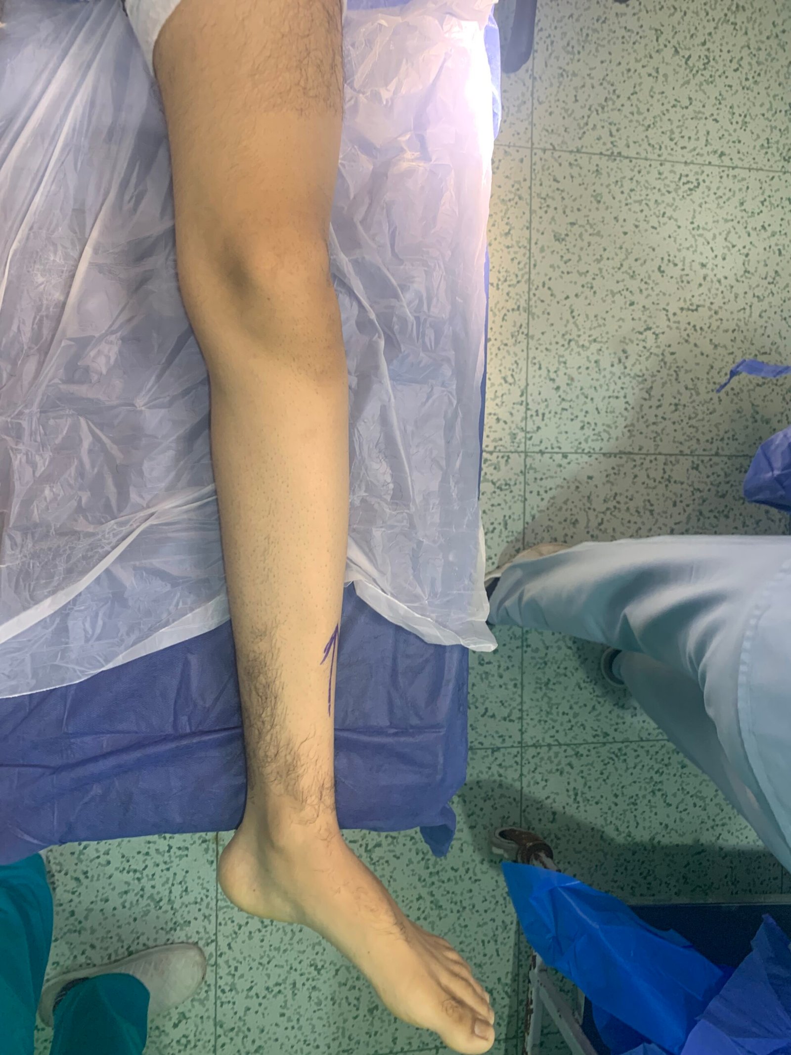
Preoperative preparation and marking.
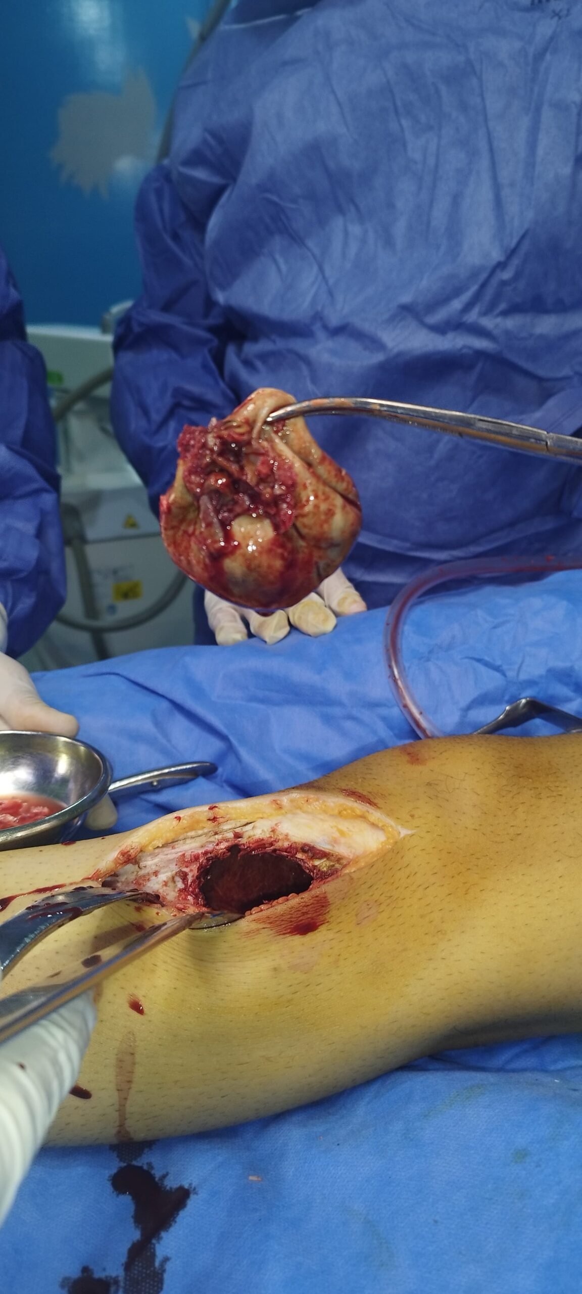
Gross specimen of the resected tumor mass.
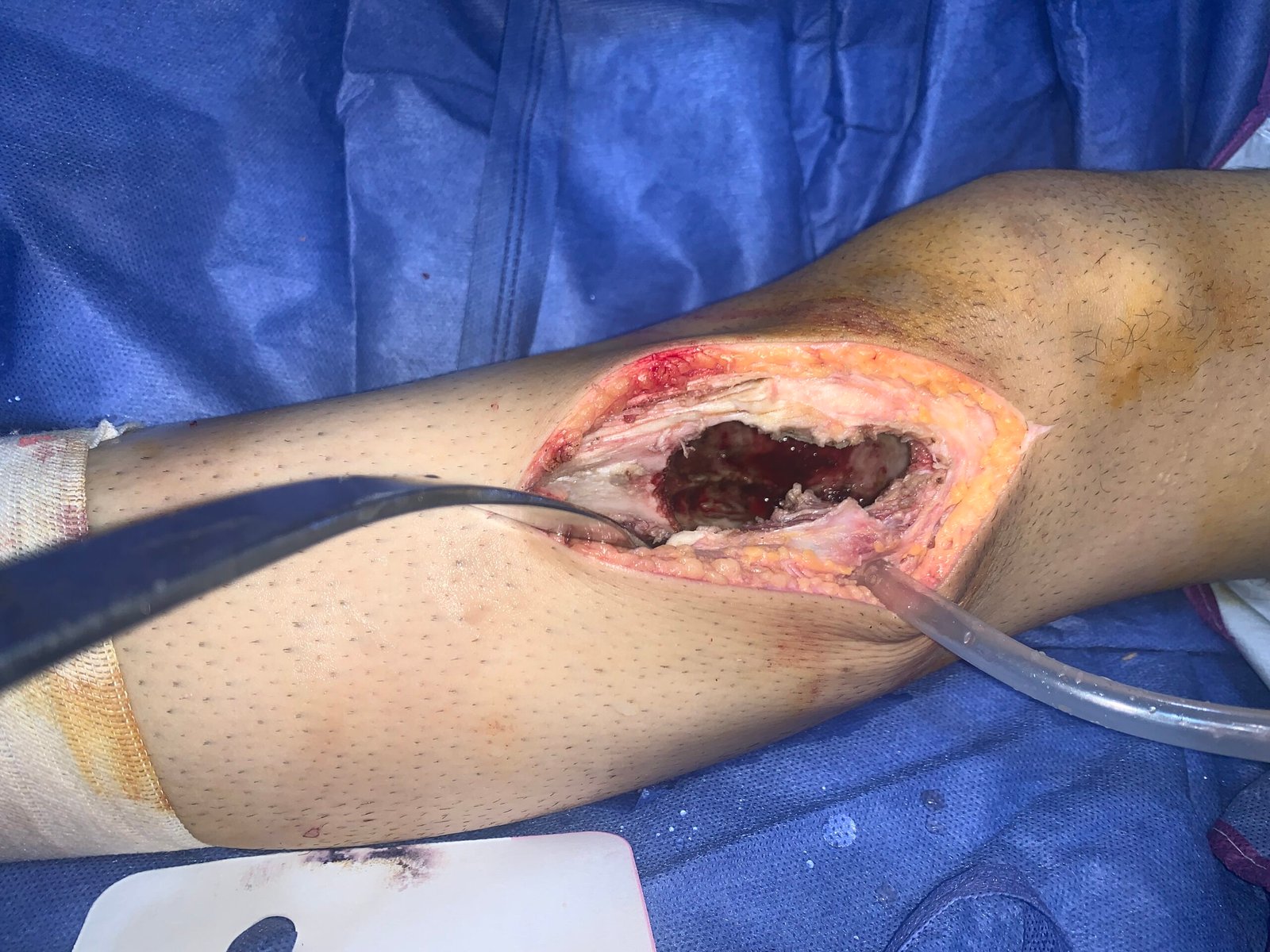
Resulting cavitary defect after initial gross curettage.
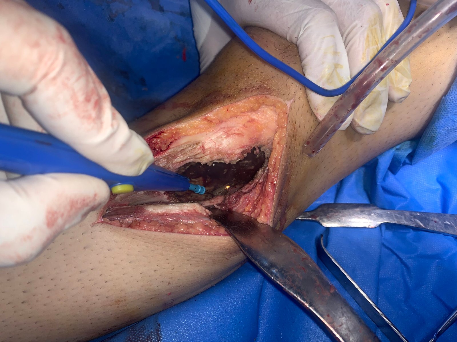
Meticulous extended curettage to clear microscopic margins.
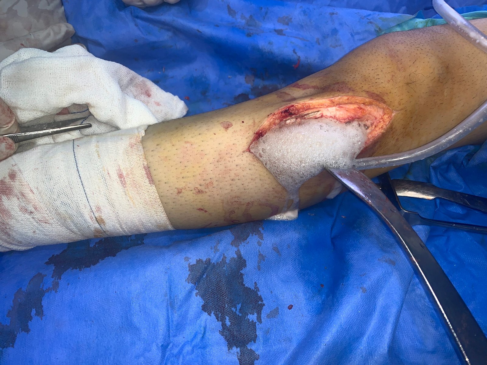
Application of thermal/chemical adjuvants for local control.

Thorough cavity wash and chemical sterilization.
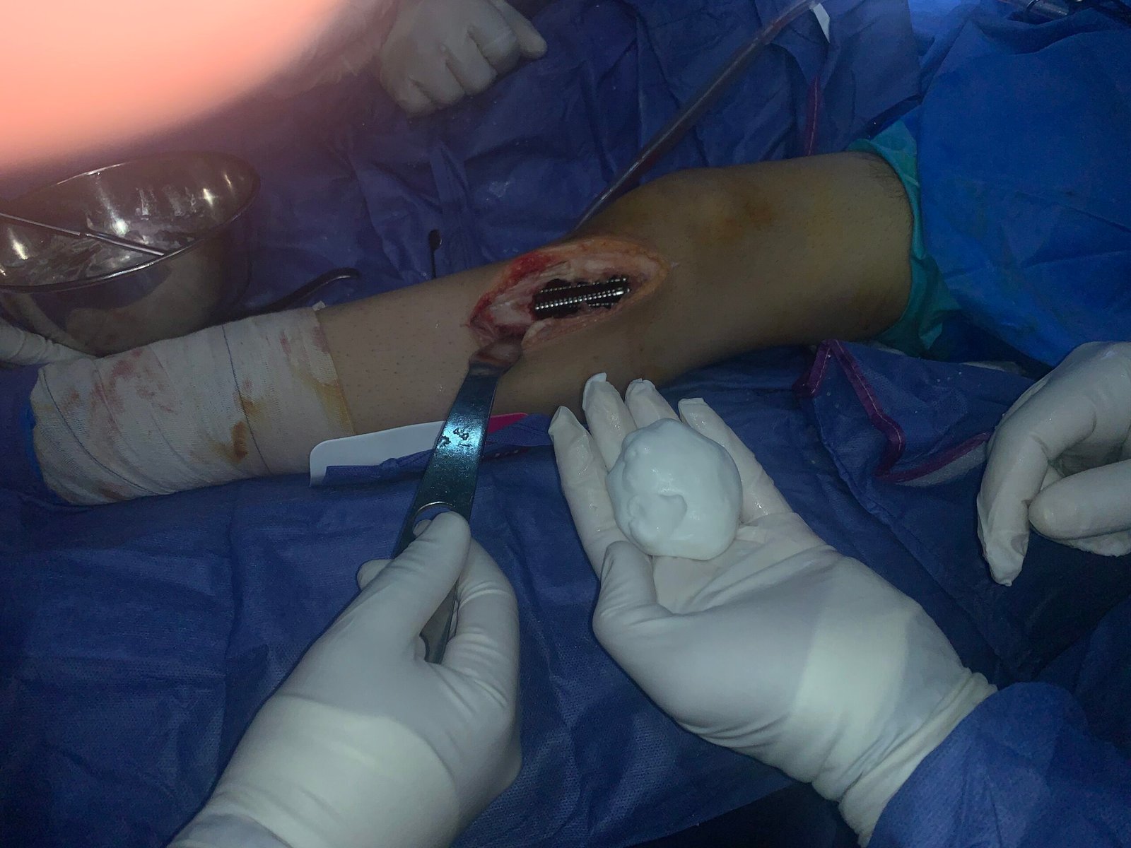
Preparation for biological or mechanical reconstruction.
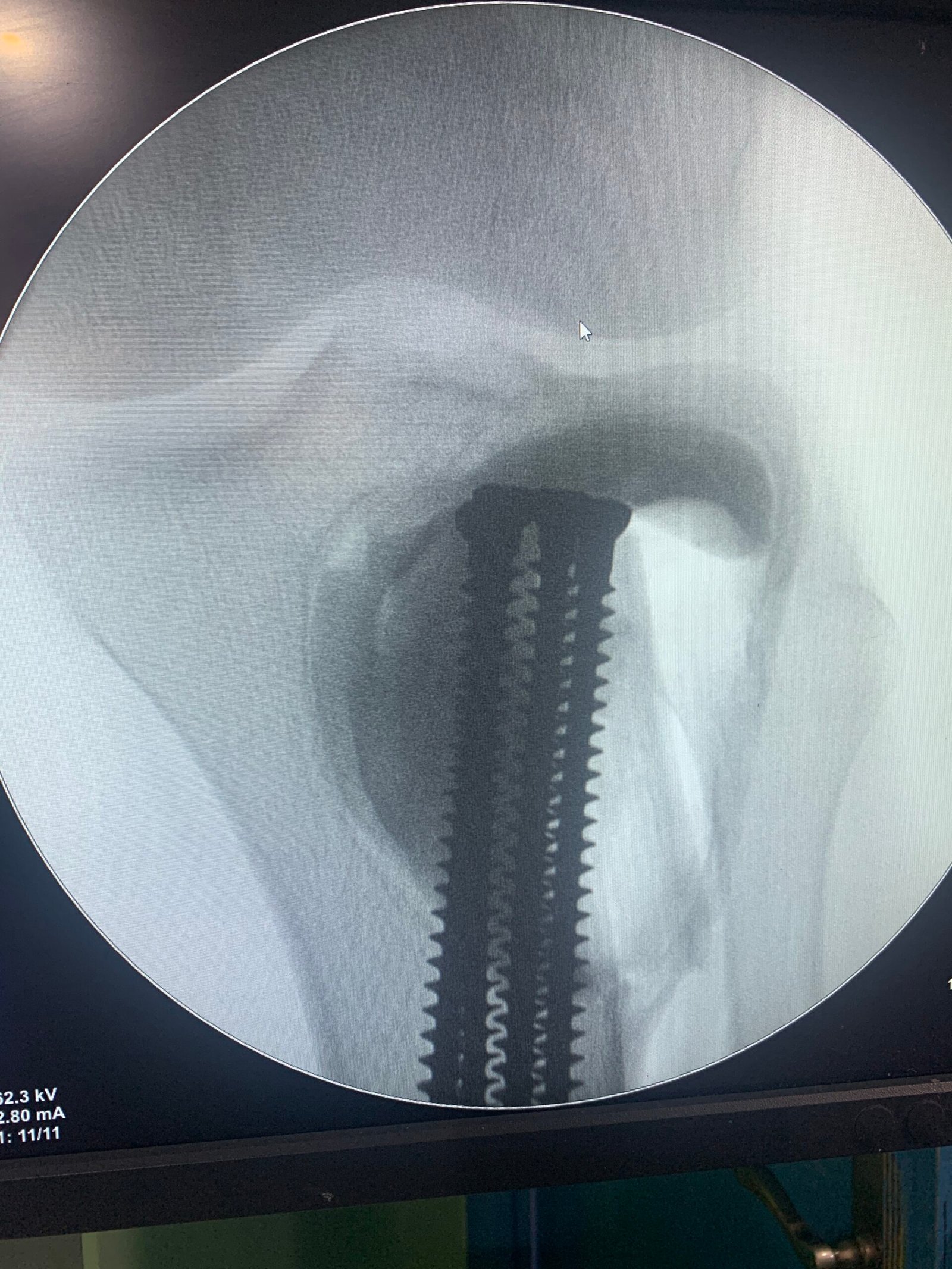
Insertion of internal fixation to stabilize the weakened cortex.
Fluoroscopic confirmation of implant trajectory.
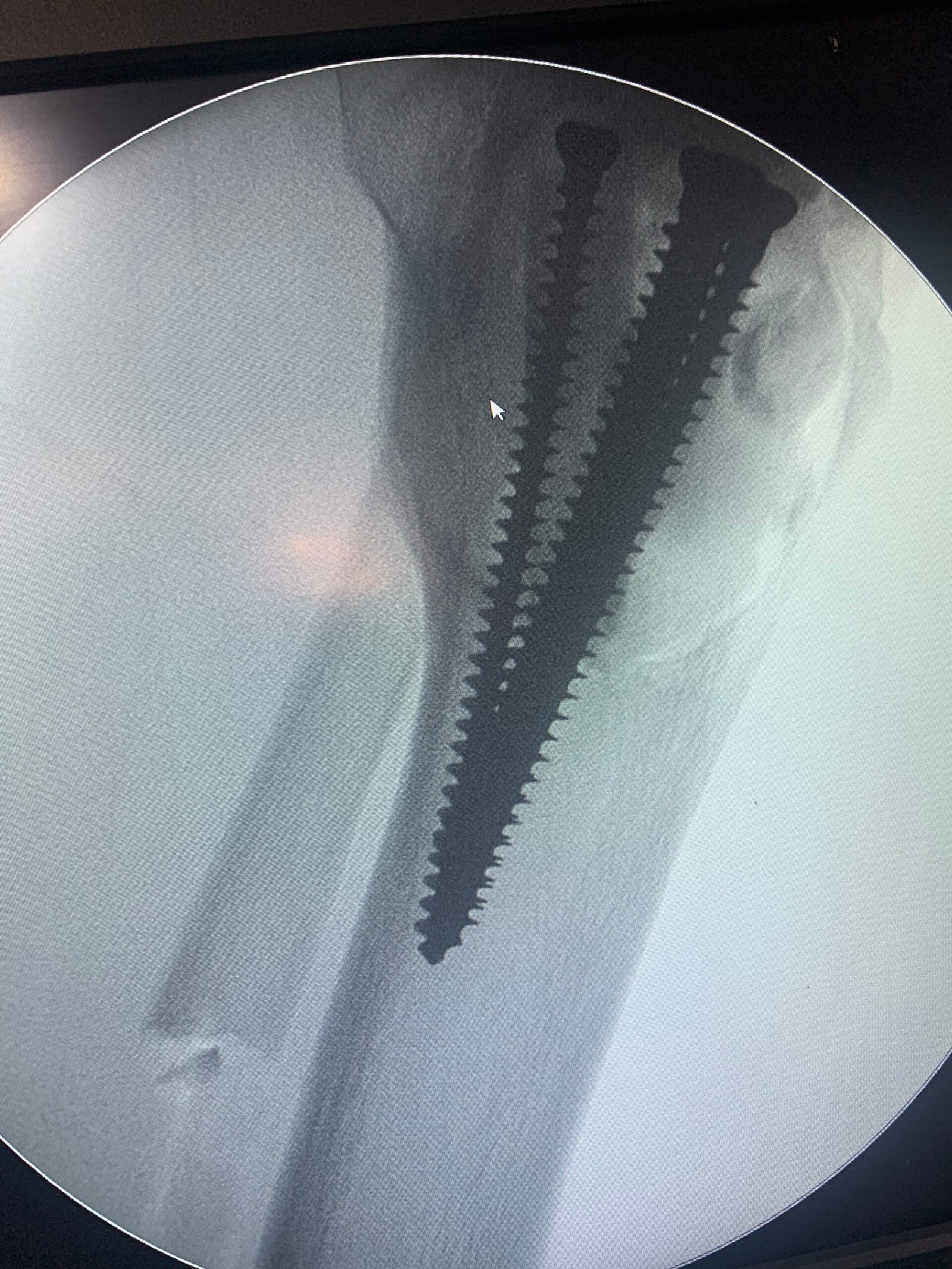
Final fluoroscopy confirming structural integrity and defect filling.
Surgical Challenges & Management Strategy
Giant Cell Tumors of bone present a unique challenge: while histologically benign, their aggressive osteolytic nature causes severe bone destruction, often extending right up to the subchondral bone of major joints. The primary goal is twofold: eradicate the tumor to prevent local recurrence while meticulously preserving the adjacent joint function.
1. The Principle of Extended Curettage
Simple “scooping out” of the tumor is insufficient, carrying a recurrence rate of up to 50%. Dr. Shaarawi employs extended intralesional curettage. After gross removal of the tumor mass, a high-speed burr is used to systematically widen the cavity by 2-3 millimeters in all directions, breaking down the bony ridges where microscopic tumor cells hide. This mechanical expansion is followed by chemical or thermal adjuvants (like phenol, hydrogen peroxide, or argon beam coagulation) to achieve a sterile, tumor-free bed.
2. Structural Reconstruction & Joint Preservation
Creating a large void in the end of a weight-bearing bone creates an immediate risk of a pathological fracture or joint collapse. To reconstruct the defect, structural support is essential. Depending on the proximity to the joint cartilage and patient factors, the cavity is filled with Poly-Methyl Methacrylate (PMMA – bone cement) or bone grafts. PMMA offers immediate structural stability and the exothermic reaction during its curing process provides an additional thermal kill zone for any residual cells. Often, internal fixation (screws or plates) is added to bridge the defect and reinforce the fragile remaining cortical shell.
Key Clinical Learning Points
The success of GCTB surgery hinges on the thoroughness of the extended curettage. Adjuvant therapies (mechanical, thermal, chemical) drastically reduce recurrence rates from >40% to approximately 10-15%.
Immediate reconstruction using bone cement (PMMA) allows for early mobilization and weight-bearing. It also facilitates easier radiographic follow-up, as any radiolucent halo developing around the radiopaque cement can signal early recurrence.
Frequently Asked Questions (GCTB Management)
Is a Giant Cell Tumor of Bone cancerous?
No, it is classified as a benign tumor. However, it is considered “locally aggressive.” This means it rarely spreads to other parts of the body (metastasizes), but it can severely damage the local bone and joint if left untreated.
Why use bone cement instead of natural bone graft?
Bone cement (PMMA) is often preferred for large defects because it provides immediate structural strength, allowing patients to walk sooner. The heat it generates as it hardens also helps kill residual tumor cells. Furthermore, if the tumor recurs, it is much easier to spot on an X-ray against the solid white mass of cement.
What happens if the tumor comes back?
Local recurrence happens in about 15-20% of cases, usually within the first two years. If it recurs, the treatment is typically a repeat extended curettage. In complex or recurrent cases, targeted medical therapies like Denosumab (a RANKL inhibitor) may be used to harden the tumor before surgery.
Dr. Ibrahim Shaarawi
Consultant Orthopedic Surgery & Joint Reconstruction
Have a similar case?
If you have been diagnosed with a bone lesion or tumor, timely expert evaluation is critical for joint preservation.
اقرأ الدليل الكامل بالعربية عن علاج ورم الخلايا العملاقة في العظام، وتعرّف على جراحة أورام العظام والأنسجة الرخوة لدى د. إبراهيم شعراوي.
المراجع (References)
- American Academy of Orthopaedic Surgeons (OrthoInfo). Giant Cell Tumor of Bone.
- Balke M, et al. Giant cell tumor of bone: treatment and outcome of 214 cases. J Cancer Res Clin Oncol. 2008;134(9):969-78.
- Chawla S, et al. Safety and efficacy of denosumab for adults and skeletally mature adolescents with giant cell tumour of bone: interim analysis of an open-label, parallel-group, phase 2 study. Lancet Oncol. 2013;14(9):901-8.
- van der Heijden L, et al. Giant cell tumour of bone in the denosumab era. Eur J Cancer. 2017;77:75-83.